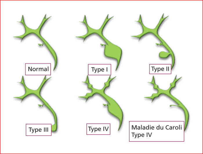
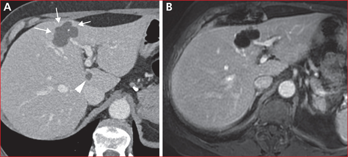
Maladie de Caroli prédominant au foie droit. Présence d’un aspect large de la voie biliaire du segment VII (flèche blanche) entourant la structure porte satellite (tête de flèche blanche).
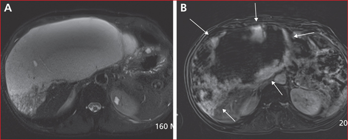
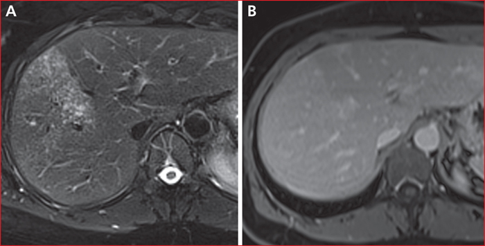
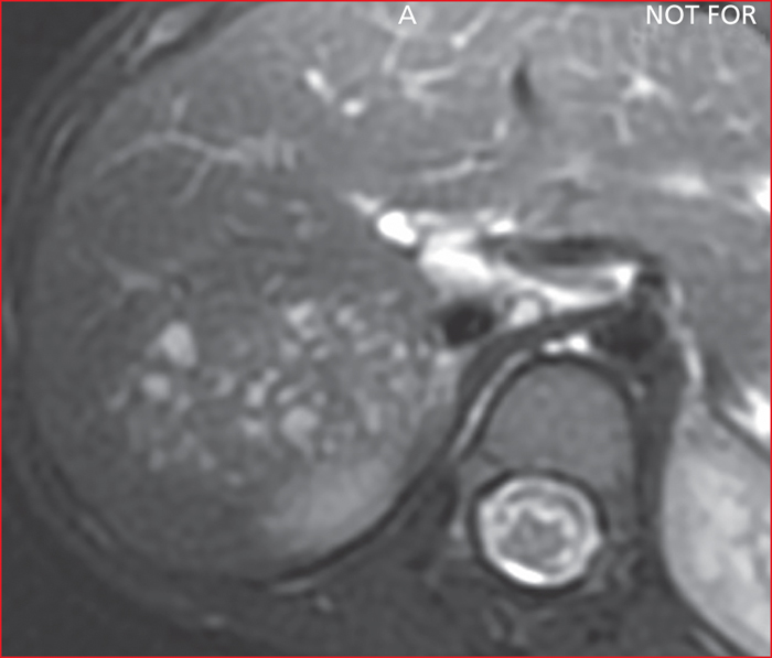
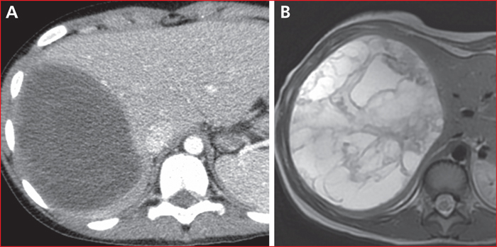
Hémangiome hépatique géant. En pondération T2, la lésion semble liquidienne si on la compare au liquide céphalo-rachidien (A). Après injection de gadolinium, il existe une prise de contraste en motte discontinue (flèches blanches), typique de l’hémangiome carverneux (B).
Péliose microkystique. La partie (A) illustre l’aspect microkystique en pondération T2. Après injection de gadolinium, les dilations sinusoïdales se remplissent permettant d’écarter des lésions réellement kystiques (B).
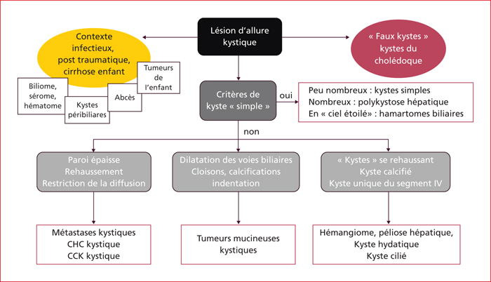
Aspect typique d’un kyste hépatique simple. En échographie (A), le kyste est trans-sonore (tête de flèche), homogène, présente une paroi fine et il existe en arrière un renforcement postérieur (flèches blanches). En tomodensitométrie (B), le kyste est hypodense (densité identique à celle de l’ascite ; têtes de flèche) et présente une paroi fine (flèche blanche). En IRM, le kyste est en hypersignal T2 (C, signal comparable au liquide, LCR ou ascite), en hyposignal T1, sans rehaussement après injection de gadolinium (D). En diffusion, le kyste est en hypersignal à b 50 (E), et en hypersignal sur la cartographie du coefficient apparent de diffusion (A, D, C, F).
Kyste simple hémorragique. La lésion est en hypersignal T1, et sa paroi se rehausse après injection de gadolinium (A). La lésion est hétérogène en diffusion (B, flèche blanche) ; à comparer avec un kyste simple non compliqué (tête de flèche).
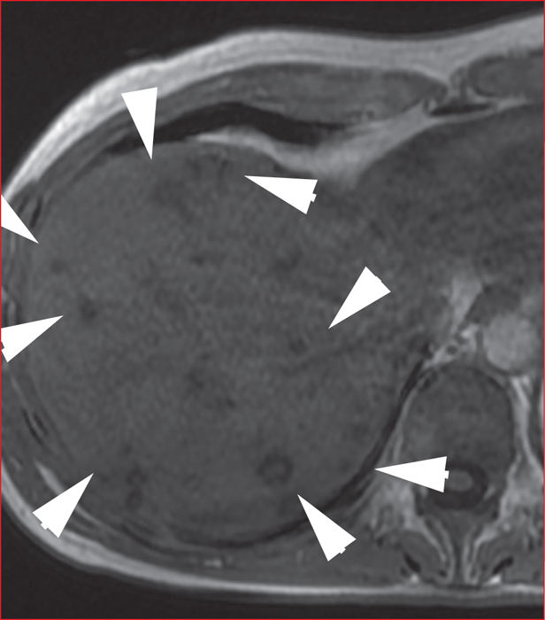
Hamartomes biliaires. Présence de multiples images kystiques en hypersignal signal T2 en IRM (A). Sur l’image de bili-IRM, la multitude de kystes est responsable de l’aspect dit de ciel étoilé (B).
Échinococcose alvéolaire en tomodensitométrie (A) et en IRM (B). Notez l’aspect kystique en tomodensitométrie, l’aspect multimicrovésiculaire en « mie de pain » en IRM, très caractéristique fait de conglomérats microkystiques sans de rétraction capsulaire. Images du Pr Valérie Laurent, CHU de Nancy France.
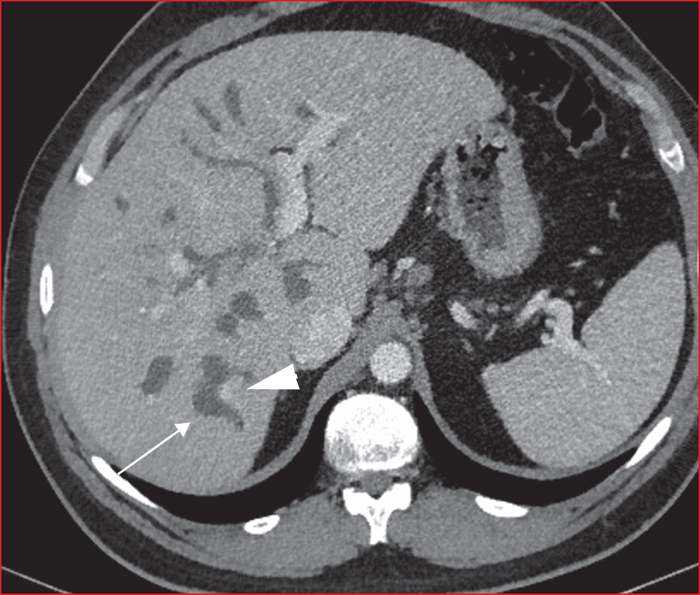
Kystes péri-biliaires dans le cas d’une cirrhose (A, tomodensitométrie, B, IRM en pondération T2). La disposition péri-portale, l’aspect en chapelet des lésions et le contexte sont évocateurs.
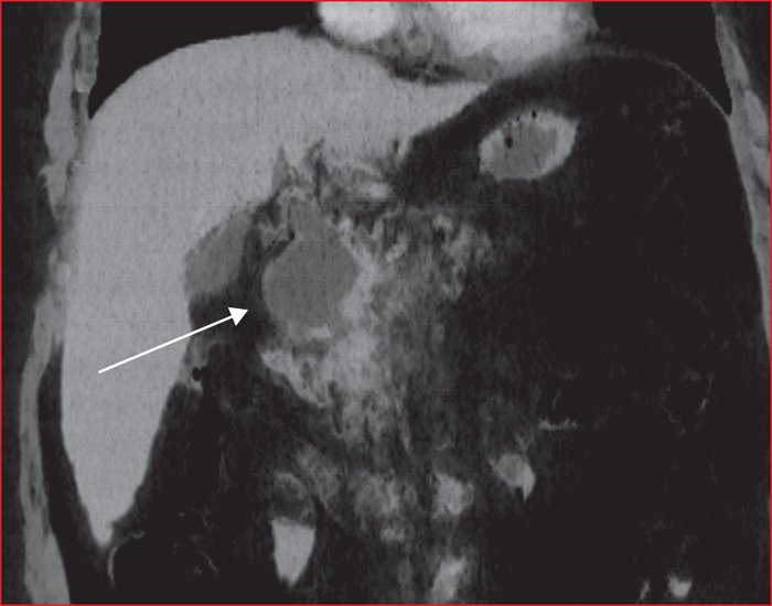
Kyste cilié. En A), aspect en tomodensitométrie et en B) en pondération T2 en IRM. La localisation du kyste, sous capsulaire et antérieure, dans le segment IV, son caractère unique sont évocateurs. Noter que la densité et le signal ne correspondent pas à de l’eau pure, comme cela serait le cas pour un kyste simple.
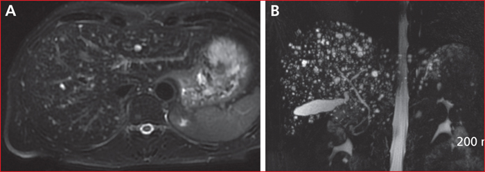
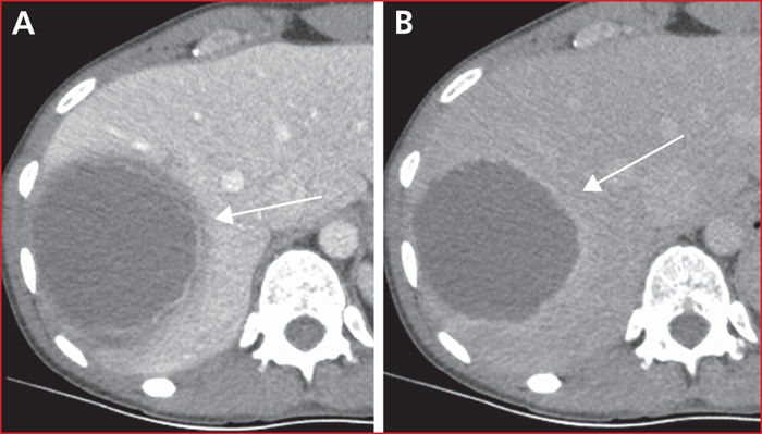
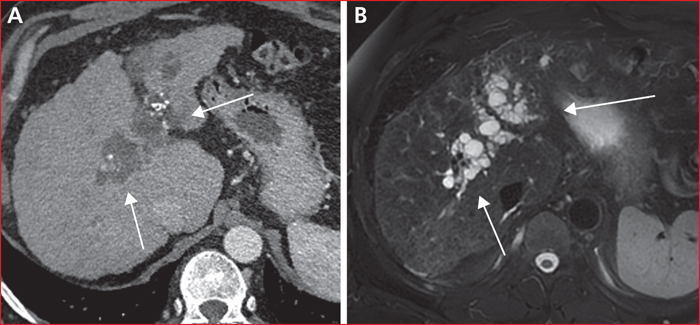
Sarcome indifférencié chez un enfant de huit ans. Noter l’aspect kystique de la lésion en tomodensitométrie (A). En IRM, la lésion est en fait très hétérogène, cloisonnée. En l’absence de contexte infectieux, le diagnostic de sarcome doit être proposé de première intention.
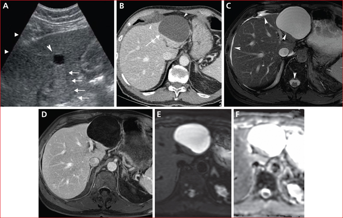
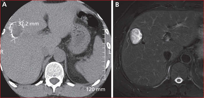
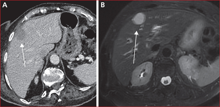
Tumeur mucineuse kystique bénigne chez une femme de 67 ans. Notez l’aspect d’intention de la lésion au niveau de l’implantation des cloisons et la calcification, peu fréquente en cas de kyste simple. A) Aspect tomodensitométrique (tête de flèche ; kyste simple du foie). B) Coupe axiale en pondération T1 après injection de gadolinium. Il existe une prise de contraste de la paroi tout à fait inhabituelle en cas de kyste simple non compliqué.
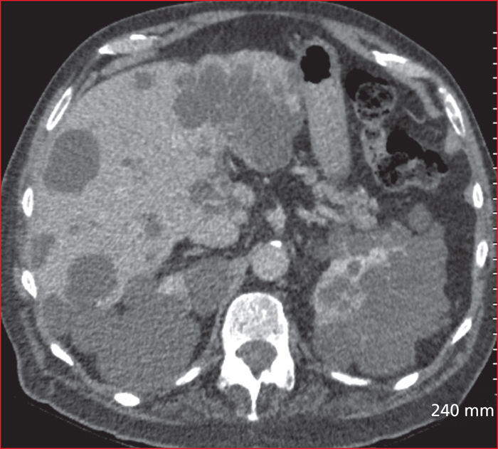
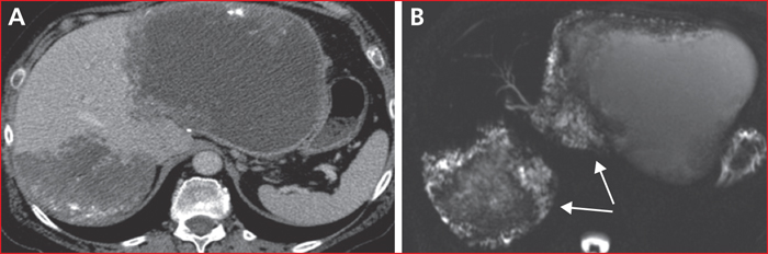
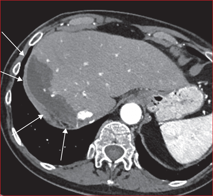
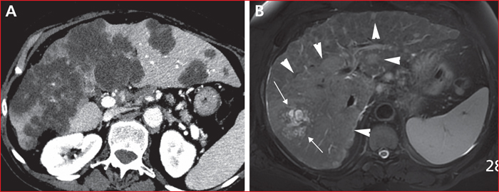
A) métastases kystique d’un cancer du côlon droit mucineux. B) Cholangiocarcinome de type masse sur une cirrhose, avec une composante kystique centrale (flèches blanches). La masse tumorale envahie une large partie du foie droit et s’accompagne de lésions filles satellites (têtes de flèche).
Benign and asymptomatic liver cysts are very frequent. This work presents the imaging features of these lesions but also other mimicking lesions.
This article describes imaging features of uncomplicated and complicated liver cysts, cystic mass of various origins, such as developmental, neoplastic or inflammatory. The diagnostic approach should take in account the clinical context, the lesion number, their dissemination and their patterns before and after contrast media injection.
A systematic approach is required to diagnose the large amont of liver cyst and a diagnostic tree is proposed to help the clinician.


 This work is licensed under a
Creative Commons Attribution-NonCommercial-NoDerivatives 4.0 International License
This work is licensed under a
Creative Commons Attribution-NonCommercial-NoDerivatives 4.0 International License