Epileptic Disorders
MENUNocturnal paroxysmal dystonia due to a subfrontal cortical dysplasia Volume 2, issue 1, Mars 2000

Introduction
Many children have abnormal motor activities during sleep. These are divided into sleep-related epileptic seizures and the parasomnias, the latter thought to be non-epileptic, developmentally-related movement disorders [2].
The term nocturnal paroxysmal dystonia (NPD) was introduced by Lugaresi et al. [3]. While still considered a parasomnia by many, it has recently been hypothesized, on circumstantial evidence, that NPD might be an epileptic disorder, arising within frontal lobe (FL) structures [4]. Others have suggested that NPD could arise from temporal [5] centro-parietal [6], lateral frontal [7] or the cingulate cortices [8]. Yet it has not been possible to localize or lateralize its epileptogenic area [4, 9-11].
While investigating childhood dyskinesias, I studied a child with NPD and demonstrated that his seizures originated from the right orbito-frontal cortex spreading to the right supplementary motor cortex (SMA). I report the cause and treatment of the NPD in this child.
Case report
1st admission
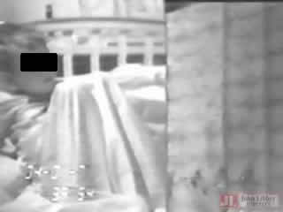
A 5-year-old boy had "frequent nightmares". His medical biography and family history were noncontributory. His general and neurological examinations (NE), EEGs and CT scan were normal. Video-EEG polygraphy captured several stereotypical events: the child aroused from slow-wave sleep (NREM) and displayed complex motor activities with dystonia of arms, trunk, neck and some ballistic movements of legs (figure 1). He appeared alert, but unresponsive. There were no automatisms or incontinence. Tachycardia and brief apneas occurred without a drop in blood O2 saturation. After about one minute, movements abated and he returned to sleep. He remembered nothing of the events. Phenytoin or carbamezapine (CBZ), afforded approximately 80% control. When he was 11 years old, he was readmitted because of relapsing frequent events.
2nd admission
The NE, interictal EEG and MRI were normal. A scalp long-term monitoring (LTM) video-EEG captured several episodes. Artifacts marred the ictal EEG. An ictal SPECT showed moderate hyperperfusion over the right frontal lobe where an interictal SPECT had revealed a moderate hypoperfusion. Since there were no localizing signs, an invasive LTM was performed (figure 2).
Twelve captured events began focally with spikes originating from the orbito-frontal cortex, spreading to the right anterior, then to the posterior interhemispheric strips recording from the supplementary motor areas (SMA), never invading fronto-parietal or temporal lobes cortices (figures 3a, b and c).
Corticography revealed spiking from a discrete area of the orbito-frontal cortex which became continuous after 25 mg of methoexital i.v. were injected. The neurosurgeon ablated 2.5 cms of this cortex.
Pathology
The surgical specimen showed features indicative of a cortical dysplasia (CD). The cortex showed no layering, neurons were of different size and orientation and some occurred in clusters (figure 4A). The white matter contained more neurons than expected at this age. Large cells, with pale cytoplasm ("balloon-like cells"), were found mainly at the border of white matter with the cortex (figure 4b).
Follow-up
There were no postsurgical deficits and, apart from two, brief convulsive events early on, the child has been asymptomatic for three years.
Horner and Jackson [12] first described attacks of "involuntary dyskinetic movements in sleep", and Lugaresi et al. studied these extensively, proposing a distinct syndrome labeled nocturnal paroxysmal dystonia (NPD) [3]. Its characteristics consisted of recurrent arousals from NREM sleep followed by dystonic, ballismic, athetoid movements. While seemingly alert, the patient would not respond. The attack terminated after two minutes or less and sleep was resumed. These authors stressed the difference between NPD and nocturnal convulsive or complex partial seizures. Although assuming a possible epileptic origin, they stressed that the evidence for epilepsy was largely circumstantial. In view of the lack of EEG abnormalities, and the striking dystonic features said to be rarely present alone in epileptic seizures, they suggested that NPD was more likely to be a type of parasomnia, namely a dystonia triggered by arousal and drew comparison with paroxysmal kinesigenic dystonia. Likewise, the 1990 classification of sleep disorders included NPD within the parasomnias [1]. Other authors also concluded that this condition could not be an epileptic disorder [13-15], some stressing the evidence militating in favor of a parasomnia, similar to night terrors [5, 16]. A psychogenic explanation for this condition was also advanced [17].
In contrast, as various expressions of frontal lobe (FL) epilepsy were detailed, more investigators inclined towards the epileptic hypothesis. Nine of 23 patients with NPD were found to have convulsions and, in spite of the lack of inter-ictal EEG abnormalities in most subjects, the authors still favored an "epileptic hypothesis" [9-11]. In two investigations [10, 11] monitoring patients with NPD and patients with established nocturnal epilepsy, no differences were observed in their clinical symptomatology regardless of the absence of EEG abnormalities in many patients with NPD. It was also suggested that the latter exhibited phenomenologies similar to seizures originating from the SMAs. However, while these were assumed to be the probable sites of origin for NPD [10, 11], it was admitted that either temporal or parietal lobes could be involved initially, the discharge later spreading to frontal structures [9-11]. Previous articles instead had mentioned the probable involvement of the temporal lobe [5] or of the pre-rolandic cortex [6].
A review, suggestively entitled: "epilepsy masquerading as a movement disorder" [4] contains the assertion that NPD is a FL seizure, and that while both temporal and frontal lobe seizures share dystonic components, other features differ. Because of the striking dyskinesia, the authors suggested a spread to basal ganglia [4]. This had been shown to occur in a patient with paroxysmal kinesigenic dyskinesia in whom a discharge starting in SMA rapidly spread to the caudate nuclei [18]. The hypothesis that NPD originates from SMA was shared by the Australian group which also concluded that NPD is indistinguishable from the syndrome known as autosomal dominant nocturnal frontal lobe epilepsy [19].
The hypothesis that NPD originates from SMA has been questioned. Thus, both clinical and electrographic differences were recently described between a large series of patients with SMA seizures and those in subjects with NPD [20]. Likewise, others, describing a syndrome of FL nocturnal partial epilepsy in children, supposedly originating from SMA, noted that it showed features distinct from those of NPD [21].
Recognizing the agreement in the recent literature in considering NPD as an epileptic rather than a parasomnia event, there are still uncertainties regarding the location of its trigger zone. Even those who favor the frontal lobe admit that the epileptogenic focus might reside in temporal or parietal lobes [9-11]. This uncertainty derives from ambiguities of reported neurophysiological and neuroimaging findings. In the few NPD patients with EEG abnormalities, these have been reported as being bilateral or diffuse, both inter and ictally. The SPECT and PET scans have also been ambiguous: some pointing to temporal or frontal lobes, others to the parietal lobes [9-11]. To my knowledge, there has been no autopsy reports on these patients and only one article was found purporting to have located the epileptogenic focus of NPD [8]. In monitoring a patient with intracerebral electrodes, his seizures were said to originate within the cingulate gyrus. Unfortunately the published iconography lacks persuasion, as indicated by others [13], and the ablation of the putative cortical focus failed to improve the patient's attacks [8].
CONCLUSION
The results of the investigations on our patient clearly support the epileptic nature of NPD, though still officially classified within the parasomnias. They also confirm its frontal lobe origin, clearly demonstrated by the invasive LTM and by the favorable results after ablation of the epileptogenic area within the orbito-frontal cortex, in this case an unsuspected cortical dysplasia (CD).
The latter location also confirms our opinion that the clinical phenomenology of the attacks exhibited by this child, mimicking those of NPD, is more like the seizures that arise from the subfrontal cortex, rather than those arising from SMAs, which had been suggested as the likely origin of NPD [4, 10, 11, 19]. Three patients with daytime seizures with a somewhat similar phenotype as those occurring at night in our child, were described by Tharp who also posited an "orbital frontal" origin for their attacks [22]. The lack of a positive familial background in this child, as in the majority of patients with NPD, suggests that many do not fit into the syndrome of autosomal dominant form of frontal lobe epilepsy with mutations in the nicotinic acetylcholine receptor subunit 4 (CHRNA4) gene. This seems even less likely for those subjects harboring a lesion, such as the CD found in this child. In a recent report on 100 patients with nocturnal frontal lobe epilepsy, only one quarter had a family history of epilepsy and none carried the CHRNA4 mutation [23], indicating the genetic heterogeneity for the ADNFL syndrome as already mentioned [19].
It also seems doubtful that the term of nocturnal paroxysmal dystonia should be maintained since it tends to imply a movement disorder or parasomnia [1] rather than an epileptic one.