Epileptic Disorders
MENULingual epilepsia partialis continua: a detailed video-EEG and neuroimaging study Volume 22, issue 4, August 2020

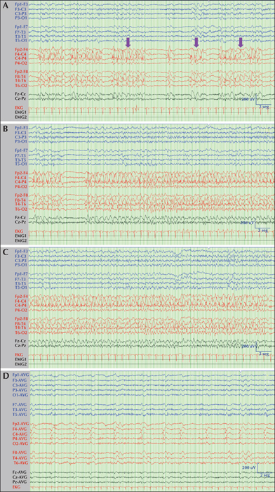
Figure 1
Ictal and interictal v-EEG features during lingual EPC. (A) Note the occurrence of lateralized periodic discharges with superimposed rhythmic activity (LPDs-plus) that occurred every two seconds with stereotyped appearance affecting a wide area of the right hemisphere. These periodic discharges conformed diplopleds and polypleds (arrows). (B, C) One of the recurrent partial motor seizures captured during the V-EEG; the patient clinically experienced tongue jerks and anarthria. (D) Note the existence of LPDs proper occurring every two seconds, involving the right frontal and temporal lobes. Low filter: 0.53 Hz; high filter: 70 Hz; notch filter: 50 Hz. vertical bar: 200 μV. Distance between solid vertical dark lines: 1 second (speed: 30 mm/second).

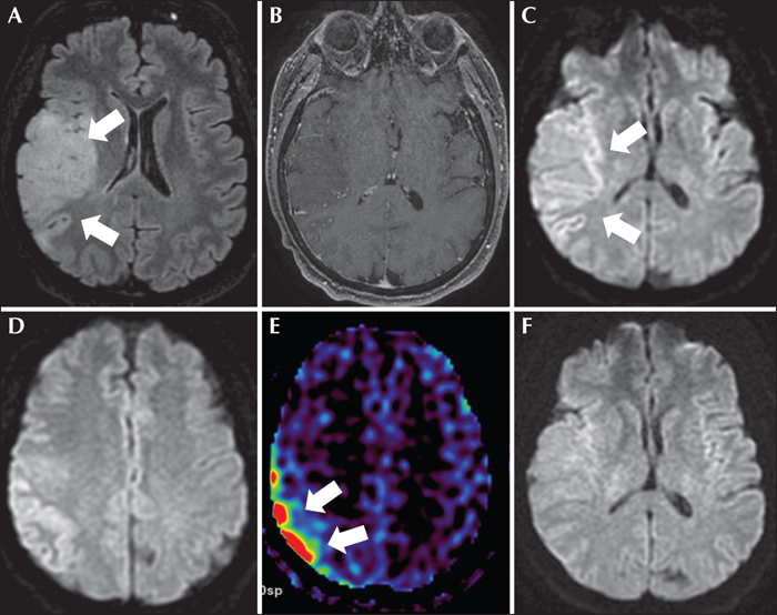
Figure 2
Brain MRI showing a right temporal cortico-subcortical lesion, hyperintense on FLAIR (A) without enhancement on T1, plus gadolinium (B), suggestive of low-grade glioma. DWI series showing restricted diffusion in the right temporo-parietal cortex (C, D) and increased cerebral flow detected on ASL series (E) suggestive of changes related to persistent status epilepticus. A second brain MRI scan was performed one month later showing persistence of the tumour (not shown) but without restricted diffusion (F) or increased blood flow (not shown).

