Epileptic Disorders
MENUEpigenetics explained: a topic “primer” for the epilepsy community by the ILAE Genetics/Epigenetics Task Force Volume 22, issue 2, April 2020

Figure 1
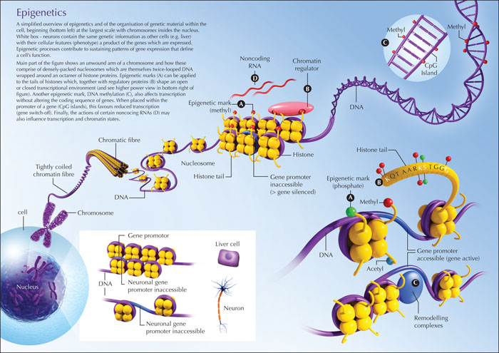
Overview of epigenetic mechanisms. A simplified overview of epigenetics and organisation of genetic material within the cell, beginning (bottom left) at the largest scale with chromosomes inside the nucleus. The white box shows neurons containing the same genetic information as other cells (e.g. in the liver) with their respective cellular features (phenotype); a product of the genes which are expressed. Epigenetic processes contribute to sustaining patterns of gene expression that define a cell's function. The main part of the figure shows an unwound arm of a chromosome and how this is comprised of densely packed nucleosomes which are themselves twice-looped DNA wrapped around an octamer of histone proteins. Epigenetic marks (A) can be applied to the tails of histones which, together with regulatory proteins (B) shape an open or closed transcriptional environment (see higher power view in bottom right of figure). Another epigenetic mark, DNA methylation (C), also affects transcription without altering the coding sequence of genes. When placed within the promoter of a gene (CpG islands), this favours reduced transcription (gene switch-off). Finally, the actions of certain non-coding RNAs may also influence transcription and chromatin states. Finally, the actions of certain noncoding RNAs (D) may also influence transcription and chromatin states.

Figure 2
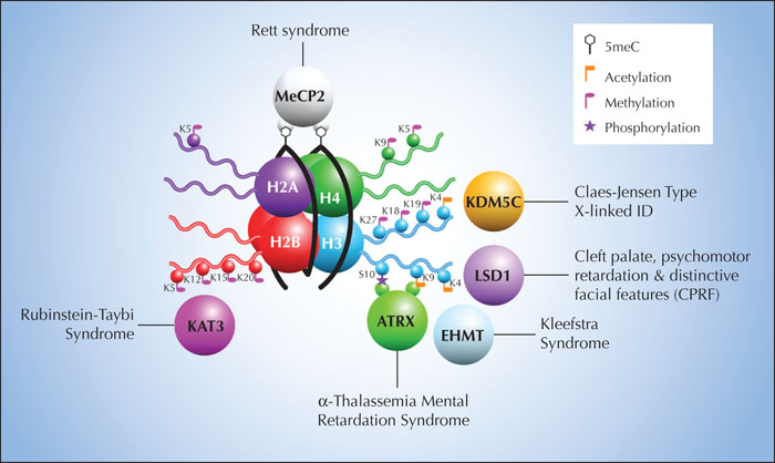
Examples of novel genes linked to epilepsy and their epigenetic functions. An example of the close relationship between chromatin function and proteins, that are mutated in patients with known syndromes often associated with intellectual disability and epilepsy, is presented. The figure shows the nucleosome, comprising a histone core and DNA coiled around it (black lines). Indicated on the histones and DNA are epigenetic marks including post-translational modifications which affect compaction and transcriptional access. Surrounding this are six proteins and their potential physical interaction sites and the known diseases caused by mutations in these. Adapted, with permission, from Iwase et al (Iwase et al., 2017).

