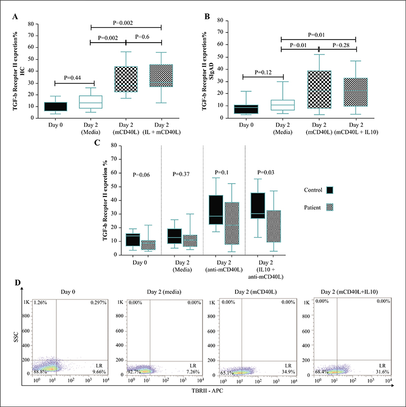
Expression of TBRII on B cells in controls (A) and patients (B) before stimulation and after culture in the presence of mCD40L alone, media or a combination of mCD40L and IL-10 for 24 h. (C) Comparison of TBRII expression in controls and patients before stimulation and after culture in the presence of mCD40L alone, media or a combination of mCD40L and IL-10 for 24 h. (D) Representative flow cytometric pattern for TBRII expression from patient data (LR: lower right).
Concentration of TGF-β secretion by B cells in control (A) and patients (B) after cultured in the presence of mCD40L alone, media or a combination of mCD40L and IL-10 for 24 h. Comparison of TGF-β secretion by B cells in study groups when treated with mCD40L and IL-10 (C).
(A) Comparison of serum TGF-β levels in patients and controls. (B) Effect of IL-10 concentration on TBRII expression and TGF-b secretion was evaluated in three control cases. Isolated B lymphocytes cultured in the presence of mCD40L (300 ng/mL) and IL-10 (20, 50 and 100 ng/mL) for 24 h. At high concentrations of IL-10, TBRII expression was decreased (red line) and TGF-β secretion (green line) was not altered.
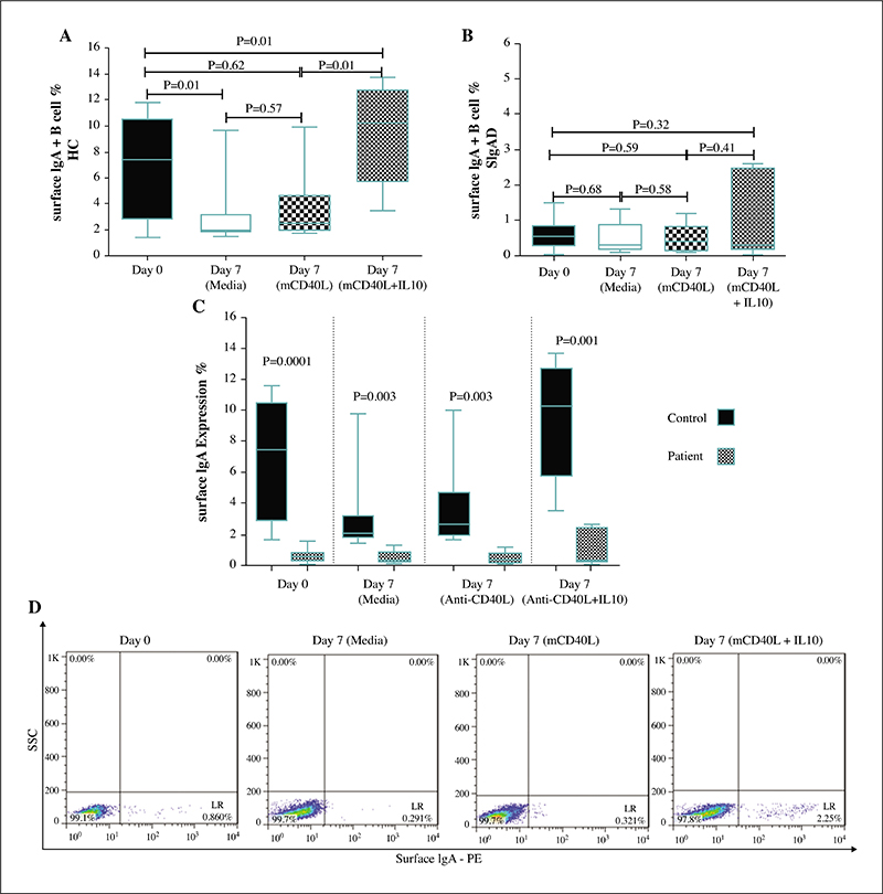
Percentage of IgA+ B cell differentiation in control (A) and patients (B) in isolated B lymphocytes before stimulation and after culture in the presence of mCD40L alone, media or a combination of mCD40L and IL-10 for 7 days. (C) Comparison of IgA+ B cell percentages in controls and patients before stimulation and after cultured in the presence of mCD40L alone, media or a combination of mCD40L and IL-10. (D) Representative flow cytometric pattern for detection of IgA expressing lymphocytes from patient data (LR: lower right).
The concentration of secretory IgA in controls (A) and patients (B) by isolated B cells after 7 days of stimulation with mCD40L alone, media and combination of mCD40L and IL-10 (C) Comparison of IgA secretion in patients and controls by isolated B cells after 7 days of stimulation with mCD40L alone, media or a combination of mCD40L and IL-10.
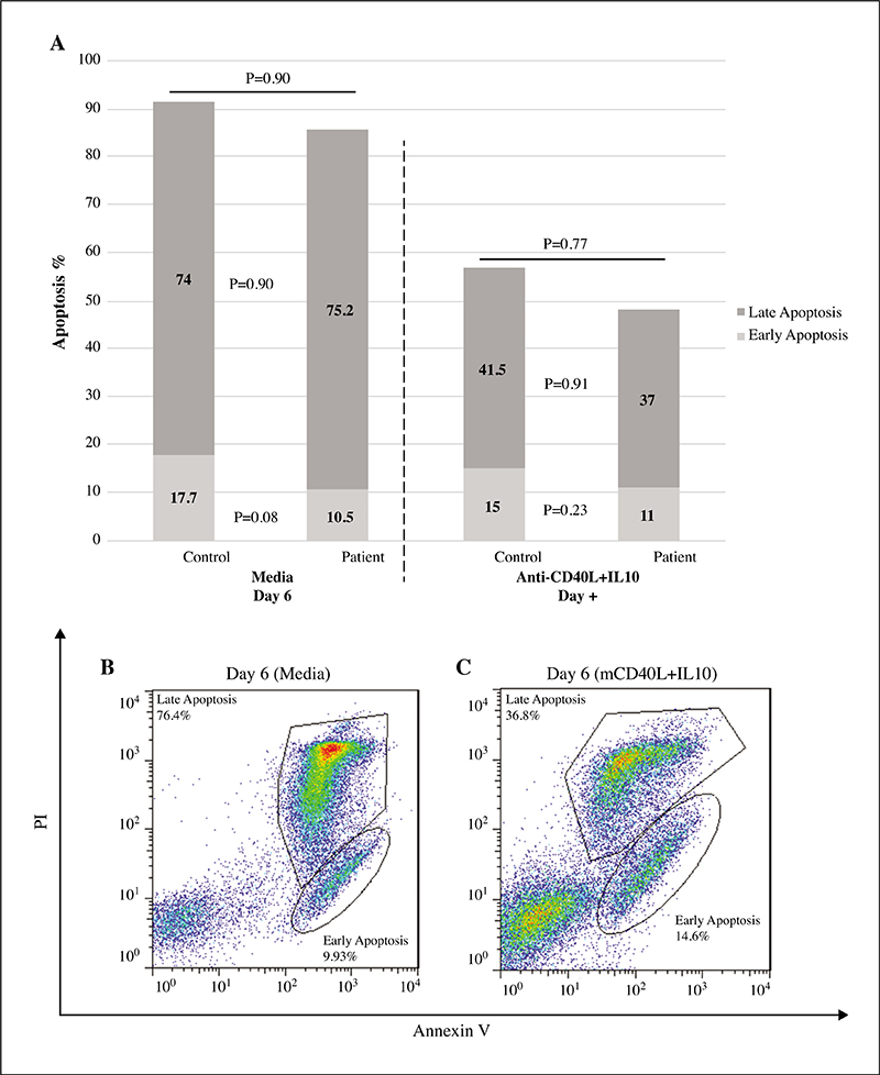
(A) The percentage of apoptosis in B cells isolated with MACS and stimulated with mCD40 + IL-10 in controls and patients after 6 days. (B) Representative flow cytometric pattern for apoptosis in unstimulated B cells and (C) stimulated B cells.
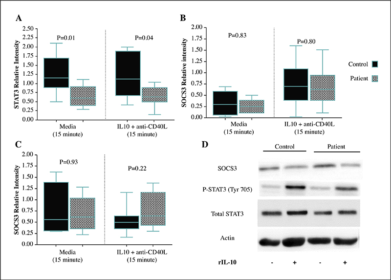
Comparison of (A) STAT, (B) p-STAT3 and (C) SOCS3 protein expression patients and controls in B cells isolated with MACS and treated with mCD40 + IL-10 in controls and SIgAD patients. Phosphorylation of STAT3 protein and expression of SOCS3 did not show a significant difference in patients compared with controls. (D) Representative blots from one experiment. STAT3 molecular weight is 85 KD and SOCS3 molecular weight is 25 KD.
1 Stem cell research center, Golestan university of medical sciences, Gorgan, Iran
2 Immunology department, Faculty of Medicine, Golestan University of Medical Sciences, Gorgan, Iran
3 Research Center for Immunodeficiencies, Pediatrics Center of Excellence, Children’s Medical Center, Tehran University of Medical Sciences, Tehran, Iran
4 Department of Neurology, Thomas Jefferson University, Philadelphia, Pennsylvania, USA
5 Immunology Research Center, Iran University of Medical Sciences, Tehran, Iran
6 Department of Immunology, School of Medicine, Iran University of Medical Sciences, Tehran, Iran
7 Non-Communicable Diseases Research Center, Alborz University of Medical Sciences, Karaj, Iran
8 Clinical Research Development Unit (CRDU), Sayad Shirazi Hospital, Golestan University of Medical Sciences, Gorgan, Iran
9 Division of Clinical Immunology, Department of Laboratory Medicine, Karolinska Institute at Karolinska University Hospital Huddinge, Stockholm, Sweden
Correspondence: Asghar Aghamohammadi, Children’s Medical Center Hospital, 62 Qarib St., Keshavarz Blvd., Tehran 14194, Iran.
Mehdi Shekarabi, Department of Immunology, School of Medicine, Iran University of Medical Sciences, Shahid Hemmat Highway, Tehran, 1449614535, Iran: M. Shekarabi
Selective IgA deficiency is the most prevalent form of primary immunodeficiencies. The pathogenesis of the disease is still unknown. Several studies have suggested a defect in B cell responses to IL-10; however, the main reason for this defect has not been reported. Elucidating IL-10 signaling defects and their correlation with clinical manifestations could be helpful for better understanding and treatment of the disease.
Methods
In this study, 15 SIgAD patients and 15 age- and sex-matched healthy controls were included. Surface expression of transforming growth factor β receptor II (TGF-β RII), IL-10R and IgA was assessed by flow cytometry in human purified B cells before and after stimulation by IL-10. Protein expression of STAT3, p-STAT3 and SOCS3 was measured by Western blotting analysis. TGF-β and IgA secretion was evaluated by ELISA. Finally, the measurement of B cell apoptosis was performed by flow cytometry.
Results
The TGF-βRII expression level was decreased after stimulation with IL-10 in patients compared with controls. Notably, the TGF-β level were higher after stimulation with mCD40L and IL-10 in the control group as compared to stimulation with mCD40L alone. The IgA+ B cell percentage and IgA secretion levels were significantly increased in controls as compared with SIgAD patients. The relative concentration of the total STAT3 was decreased as compared with controls.
Conclusion
The defect in IgA production in SIgAD patients could be due to inadequate B cell responses to IL-10 stimulation that probably originate from defective regulation of IL-10-mediated TGF-b ’symbol’ production TGF-β response by IL-10. Furthermore, it is suggested that the absence of STAT3 protein baseline expression could impair cytokine-mediated signaling such as thatinduced by IL-!0 and IL-21.