Annales de Biologie Clinique
MENUClinical value of CEA for detection of distant metastases in newly diagnosed breast cancer: comparison with CA 15-3 Volume 75, issue 4, Juillet-Août 2017

- Key words: CEA, CA 15-3, breast cancer, imaging, staging, false positive
- DOI : 10.1684/abc.2017.1258
- Page(s) : 431-41
- Published in: 2017
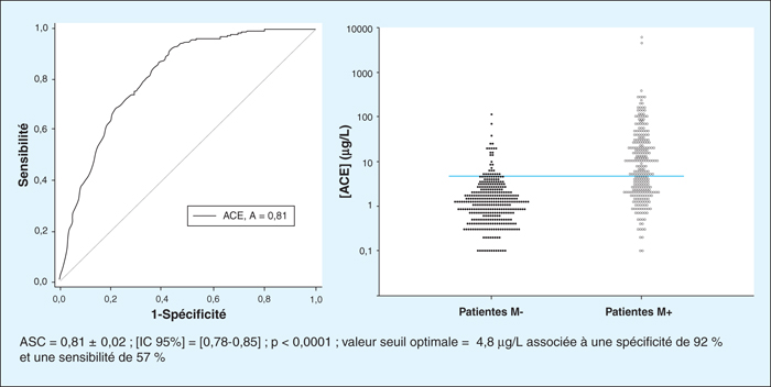
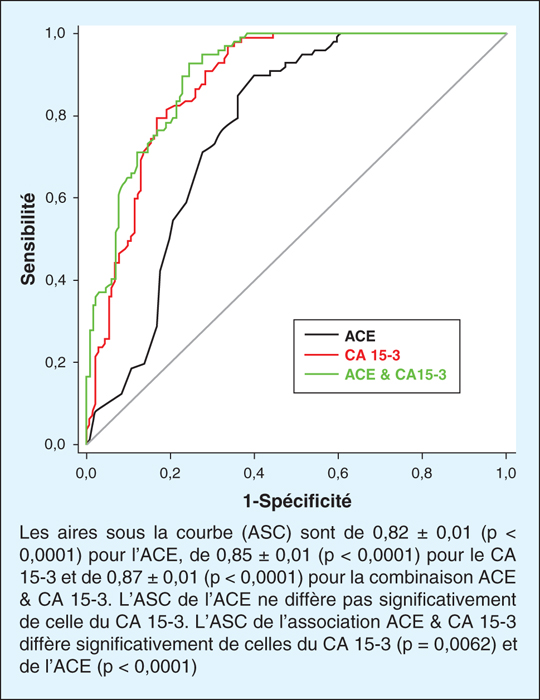
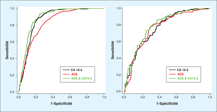
The aim of the study is to investigate the clinical value of CEA (carcinoembryonic antigen) for detection of distant metastases (DM) in newly diagnosed breast cancer. This retrospective study focuses on a population of 929 patients with locally advanced breast cancer (n=521) or metastatic breast cancer (n=408) diagnosed at the CGFL (Centre Georges Francois Leclerc) from 1998 to 2014. These patients underwent a measurement of CA 15-3, a measurement of CEA and an assessment of extension before any treatment. The initial concentrations of CEA are correlated with conventional prognostic factors. The cut-off value of CEA was determined and verified on two independents subpopulations determined in drawing lots. The ROC curve shows an AUC of 0.82 (p<0.0001). At the threshold of 6.7 μg/L, CEA before treatment has a predictive value on the existence of DM independently of the CA 15-3 and other prognostic factors. The combination of CEA and CA 15-3 increases significantly the predictive value of CA 15-3 on the whole population (sensitivity increased by 9%) and on tumors expressing hormonal receptors. Concerning the only CEA the rate of false negative is of 52% and depends on the number and the type of the metastatic localization. Among the 28 patients without DM and a CEA > 6.7 μg/L, 15 have developed DM and 2 a new cancer. Thirteen will die of cancer. In conclusion, these facts confirm the independently predictive value of CEA before treatment on the existence of DM and its complementarity with CA 15-3 during the assessment of extension by imagery.