Hépato-Gastro & Oncologie Digestive
MENUImaging of the peritoneum Volume 21, issue 8, Octobre 2014

Université Claude-Bernard Lyon 1,
service d’imagerie médicale,
165, chemin du Grand-Revoyet,
69495 Pierre-Bénite,
France
- Key words: peritoneum, computed tomography, magnetic resonance imaging, positron emission tomography, ascites, peritoneal carcinomatosis
- DOI : 10.1684/hpg.2014.1065
- Page(s) : 610-9
- Published in: 2014
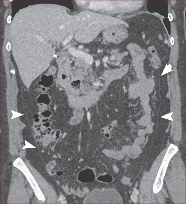
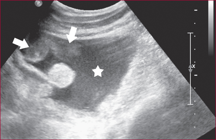
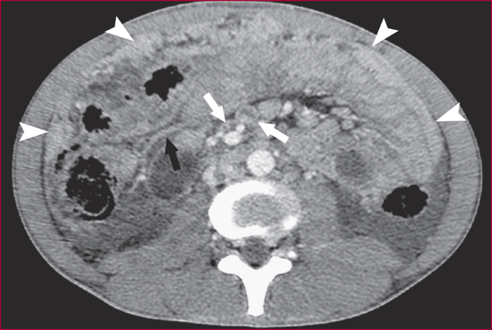
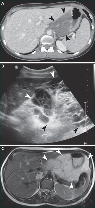
Peritoneal diseases are frequent and varied, isolated or diffused, benign or malignant, primary or secondary. Computed tomography (CT) is most commonly the first line of imaging and is currently the imaging technique reference. Ultrasonography is sometimes useful to better characterize cystic lesions when the lesions are readily accessible. Magnetic resonance imaging (MRI) can be proposed to better characterize cystic lesions, or lesions with fibrous or hemorrhagic content. Diffusion sequences allow a more functional approach and are used to improve the peritoneal staging of malignant lesions. Positron emission tomography (PET) combined with CT is less useful in characterizing the lesions and is mostly reserved for staging of malignant lesions. Considering the clinical and laboratory data, a specific radiological analysis, which sometimes requires the combination of different techniques, provides information useful for diagnostic and therapeutic strategy.