Hématologie
MENUSialylation and thrombocytopenia Ahead of print

Figure 1
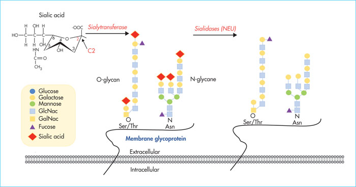
Schematic representation of the sialylation and desialylation process on glycoproteins. Sialic acid is positioned in the terminal position of N or O-glycans of a glycoprotein. It is linked by an osidic bond at position α2,3 or α2,6 to β-galactose (corresponding to the carbon position, C2, shown in red). The transfer to the glycosidic chain takes place via sialyltransferases, and elimination via sialidases (NEU), exposing the underlying sugar: β-galactose. GalNac: N-acetylgalactosamine; GlcNac: N-acetylglucosamine; Ser: serine; Thr: threonine; Asn: asparagine.
Représentation schématique du processus de sialylation et de désialylation sur une glycoprotéine. L’acide sialique est en position terminale des N ou O-glycanes d’une glycoprotéine. L’acide sialique est associé par une liaison osidique en position α2,3 ou α2,6 à un β-galactose (correspondant à la position des carbones, le C2 est représenté en rouge). Le transfert sur la chaîne glycosidique se fait par des sialyltransférases. L’élimination se fait par des sialidases (NEU) exposant le sucre sous-jacent : le β-galactose. GalNac : N-acétylgalactosamine ; GlcNac : N-acétylglucosamine ; Ser : sérine ; Thr : thréonine ; Asn : asparagine.

Figure 2
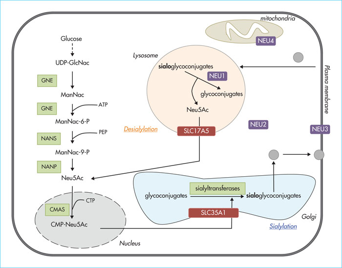
Metabolism of sialic acid in humans. Sialic acid biosynthesis begins with UDP-GlcNAc (glucose derivative) in the cytosol. Sialic acid (Neu5Ac) is synthesised in the cytoplasm as a result of the action of various enzymes. The pathway leading to thrombocytopenia involves the enzyme GNE, the transporter SLC35A1 and sialyltransferases. The free form of Neu5Ac is transformed into CMP-NeuNAc in the nucleus, and then transported to the Golgi apparatus by a specific transporter, SLC35A1, where it is transferred onto glycoconjugates by various sialyltransferases. Sialylated glycoconjugates may be desialylated in the lysosome by sialidases (NEU). Released Neu5Ac is then transferred into the cytoplasm by a specific transporter, SCL17A5, to join a new biosynthethetic cycle. The location of NEU in human cells is as follows: NEU1 = lysosome, NEU2 = cytoplasm, NEU3 = plasma membrane and NEU4 = mitochondrion. Only NEU1 and NEU2 are present in platelets. NEU1 may also be present in α granules and the plasma membrane and NEU2 in mitochondria and the plasma membrane. ATP: adenosine triphosphate; CMAS: cytosine 5′ monophosphate N-acetylneuraminic synthetase; CMP-Neu5Ac: cytosine 5′-monophosphate-N-acetylneuraminic acid; PEP: phosphoenolpyruvate; GNE: glucosamine (UDP-N-acetyl)-2-epimerase/N-acetylmannosamine; ManNac: N-acetylmannosamine; ManNac-6-P: N-acetylmannosamine-6-phosphate; ManNac-9-P: N-acetylmannosamine-9-phosphate; NANP: Neu5Ac-9-phosphate phosphatase; NANS: Neu5Ac 9-phosphate synthase; Neu5Ac: N-acetylneuraminic acid (sialic acid); UDP-GlcNac: uridine diphosphate N-acetylglucosamine.
Métabolisme de l’acide sialique chez l’Homme. La biosynthèse de l’acide sialique commence avec l’UDP-GlcNAc (dérivé du glucose) dans le cytosol. L’acide sialique (Neu5Ac) est synthétisé dans le cytoplasme suite à l’action de différentes enzymes. L’enzyme GNE, le transporteur SLC35A1 et les sialyltransférases sont impliqués dans des thrombopénies. Neu5Ac est sous forme libre puis est transformé en CMP-NeuNAc dans le noyau, et ensuite transporté dans l’appareil de Golgi par un transporteur spécifique, SLC35A1, où il sera transféré sur les glycoconjugués par diverses sialyltransférases. Les glycoconjugués sialylés peuvent être désialylés dans le lysosome par des sialidases (NEU). Neu5Ac libéré est ensuite transféré dans le cytoplasme par un transporteur spécifique, SCL17A5, pour rejoindre un nouveau cycle de biosynthèse. La localisation des NEU dans les cellules humaines est la suivante : NEU1 = lysosome, NEU2 = cytoplasme, NEU3 = membrane plasmique et NEU4 = mitochondrie. Dans la plaquette, seules NEU1 et NEU2 sont décrites. Les localisations semblent différentes avec NEU1 dans les granules α et la membrane plasmique et NEU2 dans la mitochondrie et la membrane plasmique. ATP : adénosine triphosphate. CMAS : cytosine 5′-monophosphate N-acétylneuraminique synthétase. CMP-Neu5Ac : cytosine 5′-monophosphate-acide N-acétylneuraminique. PEP : phosphoénolpyruvate. GNE : glucosamine (UDP-N-acétyl)-2-épimérase/N-acétylmannosamine. ManNac : N-acétylmannosamine. ManNac-6-P : N-acétylmannosamine-6-phosphate. ManNac-9-P : N-acétylmannosamine-9-phosphate. NANP : Neu5Ac-9-phosphate phosphatase. NANS : Neu5Ac 9-phosphate synthase. Neu5Ac : acide N-acétylneuraminique (acide sialique). UDP-GlcNac : uridine diphosphate N-acétylglucosamine.

Figure 3
Platelet life cycle. Platelets are produced in the bone marrow as a result of haematopoietic stem cell (HSC) differentiation. Megakaryocytes (MK) release platelets into the circulation. These young platelets, rich in sialic acid, will: (1) either be consumed and then eliminated by macrophages; (2) or lose their sialic acid, exposing β-galactose, to become senescent platelets. The recognition of β-galactose by AMR receptors (expressed by hepatocytes) stimulates hepatic production of TPO, allowing the formation of new platelets, and the interaction between platelets and CLEC4F, MGL and AMR, allowing their phagocytosis by Kupffer cells; macrophages present in the liver. AMR: Ashwell-Morel receptor; CLEC4F: C-type lectin domain family 4 member F; MGL: galactose macrophage type C-lectin receptor.
Cycle de vie de la plaquette. Les plaquettes sont produites dans la moelle osseuse par différenciation des cellules souches hématopoïétiques (CSH). Les mégacaryocytes (MK) libèrent les plaquettes dans la circulation. Ces plaquettes jeunes sont très riches en acide sialique. (1) Soit elles seront consommées puis éliminées par des macrophages, (2) soit elles perdront leur acide sialique, exposant ainsi le β-galactose et deviendront des plaquettes sénescentes. La reconnaissance du β-galactose par les récepteurs AMR (exprimés par les hépatocytes) stimule la production hépatique de TPO, permettant la formation de nouvelles plaquettes, et l’interaction des plaquettes avec CLEC4F, MGL et l’AMR permet leur phagocytose par les cellules de Kupffer, des macrophages présents dans le foie. AMR : Ashwell-Morel receptor ; CLEC4F : C-type lectin domain family 4 member F ; MGL : macrophage galactose type C-lectin receptor.

