Epileptic Disorders
MENUStimulus-induced rhythmic, periodic ictal discharges during funduscopic examination in a patient with status epilepticus Volume 21, issue 5, October 2019

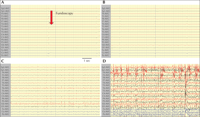
Figure 1
SIRPIDS developing during funduscopic examination on continuous EEG recording. (A) The examiner approaches the patient and starts the funduscopic examination of the patient's left eye. (B) Slightly increasing waveform changes at T6 and O2 electrodes. (C) Gradually developing higher-amplitude fast-frequency spikes, appearing at electrodes T6 and O2, without clinically overt movement. (D) Homologous and contralateral spreading of generalized spikes, which later develop into a clinical generalized tonic-clonic seizure. The EEG settings were as follows: sensitivity: 10 μV/mm; low-frequency filtre: 0.3 Hz; high-frequency filtre: 70 Hz.

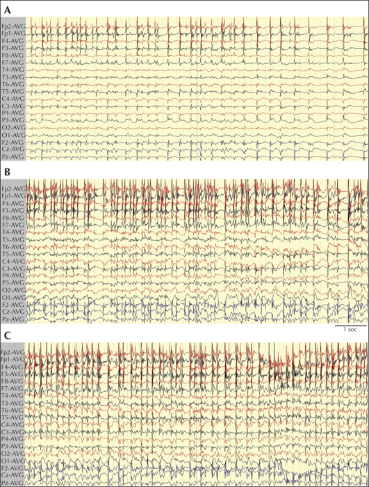
Figure 2
Interictal diffuse abnormalities not related to funduscopic stimulation. (A) Quasi-periodic high-amplitude spikes, predominantly in the right frontal area (Fp2F4F8), which occasionally evolved into quasi-periodic generalized spikes, corresponding to a clinical generalized seizure (B, C). The EEG settings were as follows: sensitivity: 10 μV/mm; low-frequency filtre: 0.3 Hz; high-frequency filtre: 70 Hz.

