Stereotactic-EEG-guided radiofrequency multiple hippocampal transection (SEEG-guided-RF-MHT) for the treatment of mesial temporal lobe epilepsy: a minimally invasive method for diagnosis and treatment
Volume 23, issue 5, October 2021
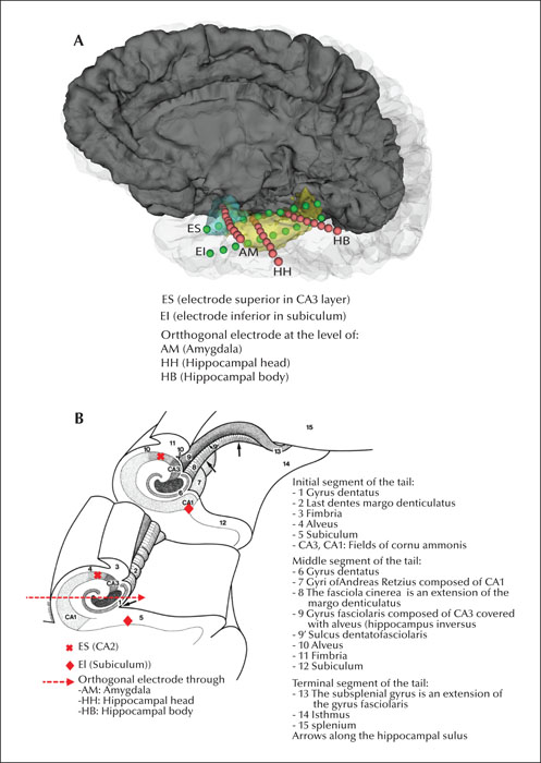
(A) Three –dimensional schematic protocol of SEEG-electrode position for mesial temporal SEEG monitoring and radiofrequency trans-ablations. Three orthogonal electrodes to the mid-plane at the level of the amygdala (AM), head of hippocampus (HH) and body of hippocampus (HB) were implanted (pink electrodes). Note the two additional electrodes with trans-occipital approach implanted along the longitudinal axis of the hippocampus (green electrodes): one implanted in the most apical area of the hippocampus (electrode superior, ES) and a second electrode (electrode inferior, EI) which was implanted parallel to ES and underneath to the amygdalohippocampal complex in an area between the subiculum, entorhinal area and the inferior longitudinal fasciculus. Also, note that electrode AM is located above ES. However, HH and HB electrodes are located between electrodes ES and EI. (B) Diagram of coronal section of the hippocampus and spatial relationship between electrodes and different structures within the hippocampus. Note the two electrodes with trans-occipital approach implanted along the longitudinal axis of the hippocampus. ES (Entorhinal Superior) is located near the alveus and fimbria within the CA2 layer. EI (Entorhinal Inferior) is located between the subiculum and the entorhinal area. The orthogonal electrode at the level of the head and body of the hippocampus (HH) is located between ES and EI. At the level of the amygdala the location of the orthogonal electrode is superior to ES and EI. Two or three radiofrequency transections can be performed using contacts of adjacent electrodes (ES-OE and OE-EI). (Diagram taken from “The Human Hippocampus. An Atlas of Applied Anatomy” Henri M. Duvernoy J.F. Bergmann Verlag Munchen. With permission from Springer Nature).
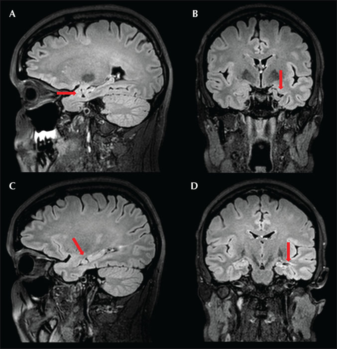
Sagittal and coronal Brain MRI (FLAIR sequence) of a patient obtained 6 months after SEEG guided-RF-MHT. The MRI shows linear thermocoagulative lesions perpendicular to the longitudinal axis of the amygdalahippocampal complex (arrows). (A) and (B) At the level of the amygdala. (C) and (D) At the level of the head of the hippocampus.
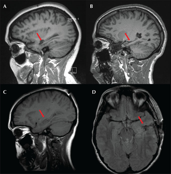
Brain MRI at 6 months follow up of three different patients who had the standard MHT technique via open craniotomy (T1 sequence MPRAGE). (A) Sagittal view MRI of a patient that underwent MHT (-) technique (without additional resections). Note, some loss of hippocampal volume associated with this technique (arrow). (B) Sagittal view MRI of a patient that had a modified MHT (+) technique. Note the temporal tip neocortical resection, amygdalectomy and greater associated hippocampal volume loss (arrow). (C) Sagittal and axial view (FLAIR sequence) brain MRI of a patient that had MHT (-) (without additional resections). Note, in this patient the adequate preservation of the hippocampal volume (arrow). However, note the damage produced to the temporal stem and neocortical area associated to the surgical approach.
1 Department of Neurology, University Hospitals Cleveland Medical Center, Cleveland, Ohio
2 Case Western Reserve University, School of Medicine, Cleveland, Ohio
3 Department of Neurosurgery, University Hospitals Cleveland Medical Center, Cleveland, Ohio
4 Neuroscience Department, Kaiser Permanente Northern California, Redwood City, California
5 The University of Texas Health Science Center at Houston, McGovern Medical School, Houston, Texas, USA
6 Epilepsy Center, UH Rainbow Babies & Children's Hospital, Case Western Reserve University School of Medicine, Cleveland, Ohio, USA
* Correspondence: Naiara Garcia Losarcos
Department of Neurology, Epilepsy Center
University Hospitals Cleveland Medical Center
11100 Euclid Avenue, Cleveland, OH 44106, USA
For the treatment of mesial temporal lobe epilepsy on the language-dominant side in patients at high risk of memory decline, we propose a minimally invasive diagnostic and treatment technique, adopting the principles of multiple hippocampal transections (MHT) using stereo-electroencephalography-guided radiofrequency (SEEG-guided-RF-MHT). This new technique allows targeting of the longitudinal fibers in the hippocampus critical for seizure spreading, while sparing the transverse circuits which are considered important for memory processing and avoiding discomfort and longer post-operatory recovery time associated with craniotomies. We report the efficacy and safety of this procedure in a preliminary observational study of cases.
Methods
Five patients at high risk of memory decline, including three with non-lesional hippocampi on MRI, had temporal lobe epilepsy (TLE) necessitating depth electrode implantation. A new strategy of SEEG electrode placement was used to mimic MHT. After confirming hippocampal seizure onset, all the patients had three linear ablations perpendicular to the amigdalohippocampal complex. The procedure was performed at the patient's bedside with the patient awake during the full length of the procedure.
Results
Four out of five patients were seizure-free (average follow up: 14-18 months). There were no associated complications. Visual inspection of brain MRI of patients at six months following SEEG-guided RF-MHT showed significant hippocampal volume preservation. Subjects who received the procedure in the dominant side reported no subjective memory complaints in the follow-up clinic assessments at six months.
Significance
Our preliminary seizure outcome seems very promising since the majority of our patients (four out of five patients) were seizure-free. Since no lesions are made outside the amygdalo-hippocampal complex using this technique and the temporal stem remains intact, more favorable memory and language outcome is expected in patients at high risk of memory decline.