Epileptic Disorders
MENUSleep-related laryngospasm: a video-polysomnographic recording Volume 8, issue 1, March 2006

Auteur(s) : Jorge Iriarte1, Elena Urrestarazu1, Manuel Alegre1, Concepción Goñi2, César Viteri1, Julio Artieda1
1Clinical Neurophysiology Section, Department of
Neurology, Clínica Universitaria, University of Navarra, Pamplona,
Navarra, Spain
2Pediatric Neurology Section, Hospital de Estella.
Navarra, Spain
Article reçu le 25 Mai 2005, accepté le 21 Novembre 2005
Sleep-related laryngospasm is a rare phenomenon, included in the diagnostic and coding manual of the American Sleep Disorders Association as a sleep disorder associated with conditions classifiable elsewhere (ASDA, 2005). Only a few references are found in the neurological literature. It is defined as a dramatic attack that produces breathing difficulties, awakening, stridor, apnoea, fear, agitation, and even exhaustion. It resolves in 1-3 minutes. The cause may be related to epilepsy (Amir et al., 1983, Cohen et al., 2000) or, more often, to gastroesophageal reflux (Campbell et al., 1990, Thurnheer et al., 1997). The mechanism includes a paroxysmal closing of the vocal cords, probably induced by a vagal response or as part of an epileptic seizure. The presence of stridor is the sign that most typically supports this diagnosis against the other possibilities.The goal of this report is to present a video-documented case of nocturnal laryngospasm with very typical semiology. This case illustrates not only the clinical findings but also the EEG and polysomnographic recordings, the aetiology, and the response to the treatment.Case study
We present the case of a 12-year-old boy who, during the previous two months, had suffered from sudden nocturnal episodes of awakening, coughing, and breathing difficulties. The episodes occurred once or twice per night, three to four days each week, starting around one hour after falling asleep. The results of the physical examination were normal. A full otolaryngological examination was performed and was found to be within normal limits. He did not suffer from any allergies or asthma. There was no family history of epilepsy, and the boy did not have any other remarkable medical history. A standard EEG was also normal. He was sent to our center for a video-polysomnographic study.He was monitored in our sleep unit using a Stellate system with the Harmonie 5.2b software (Stellate, Montreal, Canada), using digital video and EEG, and Lamont 32-Sleep amplifiers (Lamont Medical, Wisconsin, USA). The polysomnographic recording (PSG) included EEG (Fp1, Fp2, C3, C4, Cz, O1, O2), referenced to both balanced mastoids), electro-oculogram (EOG), oxygen saturation level, airflow, respiratory band, EMG in anterior tibialis, ECG and an Opti-Flex™ (Sleep Mate, Midlothian, VA, USA). To evaluate airflow, we used a pressure transducer (PTAF-2, Protech, Woodinville, WA, USA). The Opti-Flex™ sensor was designed to measure the movement in the sternal notch associated with respiratory effort; it is considered useful in disorders without full apnoeas such as the upper airway resistance syndrome. The mother gave her written consent for the procedure.
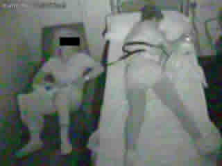
The patient had two episodes; both were very similar and occurred while he was sleeping, at 00:26 am and 5:47 am respectively. The video showed a sudden awakening with coughing, stridor, choking, and agitation, very similar to the description provided by the mother. The polysomnographic recording showed that the boy was in stage 3-4 of sleep. He started having obstructive apnoea (no flow while movement of the respiratory band is present), with a change in the suprasternal sensor (( figure 1 )). He continued experiencing intermittent apnoeas while awake. Each episode lasted almost three minutes, after which he went back to sleep. The rest of the sleep study was normal, without epileptiform activity or additional sleep apnoeas.
Apart from laryngospasm, the proposed differential diagnoses included frontal seizures, sleep-related choking syndrome, sleep-related asthma, sleep apnoea, and REM sleep behaviour disorder (RBD). The presence of stridor differentiated between laryngospasm and the sleep-related choking syndrome. The boy had not previously suffered from asthma and never suffered attacks during the day. RBD was discounted because the episodes occurred in slow sleep, (a similar case has been reported by Aloe and Thorpy, 1995). Frontal seizures may mimic the observed semiology and often exhibit a normal EEG.
The diagnosis of sleep-related laryngospasm was proposed because of the semiology. A week later, a nocturnal pH-metry was performed; it was consistent with a marked gastroesophageal reflux. During this study, the patient did not experience any clinical episodes. Under antireflux therapy, the episodes gradually became more mild and infrequent, effectively stopping after three weeks. In the last four months, he has not experienced any other episodes, and therefore the diagnosis of sleep-related laryngospasm was established. The non-epileptic nature of the events was confirmed by the positive response to this treatment.
The mechanism of the laryngospasm provoked by reflux is not clear, but a vagal mechanism has been proposed (Benhamou and Dupont, 1992). Also, as explained above, some cases of laryngospasm are due to epileptic discharges, possibly both frontal and temporal, and in fact, in 30% of cases, carbamazepine is an effective treatment. It is important to take into consideration the fact that laryngospasm may appear in the absence of any other symptoms of gastroesophageal reflux (Fonkalsrud and Ament, 1996).