Epileptic Disorders
MENUSeizure heralding tuberculous meningitis Volume 14, issue 3, September 2012

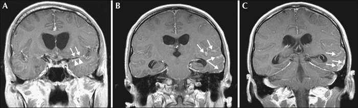
Figure 1 (A) Enhancement of the wall of the left middle cerebral artery (arrows) and upper surface of the left temporal pole (arrowheads) are noted (B) Thickening and enhancement of the subarachnoid spaces and >dura mater are noted in the left temporal lobe (arrows) Enhancement extends into the left Sylvian fissure (C) Irregular thickening of the dura mater and enhancement of the sulci in the left posterior temporal lobe are noted (arrows).
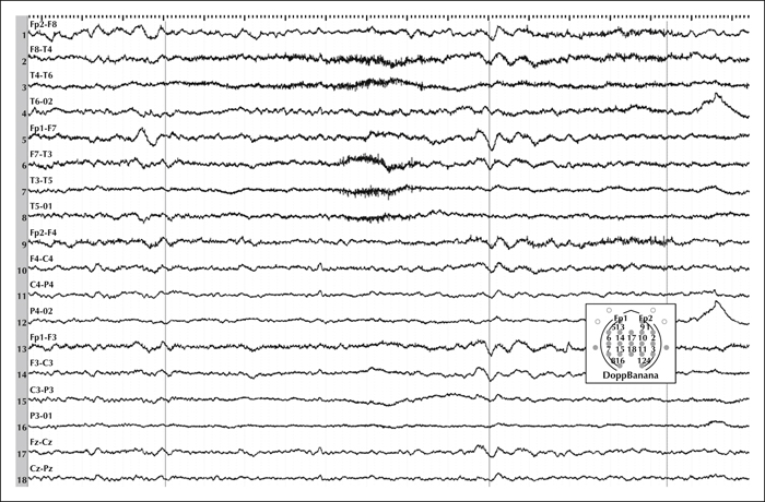
Figure 2 Pattern of diffuse EEG slow waves without clear lateralisation or evident epileptiform abnormalities. Sensitivity: 7 μV/mm; low filter: 0.53 Hz; high filter: 70.0 Hz; paper speed: 20 sec/page; each vertical bar: 1 sec.