Epileptic Disorders
MENUPilomotor seizures: a video case report Volume 14, issue 1, March 2012

epd.2012.0496
Auteur(s) : Mohamad Ayman Haykal, Bassel Abou-Khalil bassel.abou-khalil@vanderbilt.edu
Department of Neurology, Vanderbilt University Medical Center, Nashville, USA
Correspondence: Bassel Abou-Khalil 1161 21st Ave South, A-0118 Medical Center North, Nashville, TN 37232-2551, USA
Piloerection is a rare ictal manifestation that is commonly overlooked by patients. In addition, objective evidence of piloerection is seldom available due to limitations of video resolution and the impracticality of continuous exposure of involved body areas in the epilepsy monitoring unit (EMU). We documented ictal piloerection with high-resolution video recordings in a patient with frequent pilomotor seizures evaluated in the EMU. We also present video-EEG and imaging findings and discuss the localising and lateralising value of this interesting ictal phenomenon.
Case Study
A 75-year-old right-handed male was presented to the Vanderbilt Medical Center EMU for evaluation of spells of unknown nature. Approximately one year prior to admission in the EMU, he developed shingles on his right leg, then, a few days later was hospitalised for confusion and diagnosed with herpes zoster encephalitis. He made a full recovery, but seven months later started having daily recurrent episodes involving a quick jerking movement of the right arm and right facial drawing for a few seconds. Three months later, ten months after he was hospitalised for encephalitis, the patient presented with different symptoms. He would feel a hot flash starting in the head and running down his body, with “goose bumps” spreading in a similar fashion. These were most noticeable in the thighs. Occasionally, an episode was preceded by a very brief recollection of emotionally neutral remote memories with no associated fear or anxiety. He usually maintained awareness during the spell but at times his speech became slurred or was interrupted for a few seconds. Episodes lasted less than 30 seconds and occurred at least 10 times a day.
His initial evaluation included magnetic resonance imaging (MRI) of the brain and an electroencephalogram (EEG), both reportedly normal. A 24-hour video-EEG monitoring study captured the first type of spells which were deemed to be non-epileptic. Both types of spells continued despite treatment with levetiracetam and gabapentin.
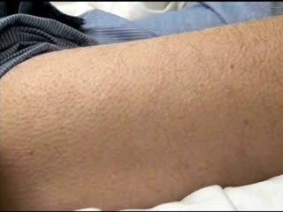
During his evaluation in our EMU, the patient had 40 events. For most events, he reported a subjective feeling of a “hot flash” starting in the head and spreading downwards, in addition to piloerection that mainly involved the bilateral lower extremities. In some seizures, there were word-finding difficulties, stuttering or dysarthria and occasionally a jaw tremor when the patient attempted to speak. A phonemic paraphasic error was recorded in one seizure. A few seizures were characterised by brief arousal from sleep, of which the patient had no recollection. Corresponding to these events, the EEG showed an ictal discharge of theta range with consistent left inferomesial-anterior temporal onset (figure 1). Interictal epileptiform discharges had a similar distribution (figure 2). Piloerection was objectively demonstrated on video (see video sequence 1); the onset of piloerection preceded the onset of ictal discharge on scalp EEG by 15 seconds.
Subsequent to his EMU evaluation, the patient underwent MRI of the brain which showed mild T2 hyperintensity in the left mesial temporal lobe, without associated volume loss (figure 3). Positron emission tomography (PET) of the brain showed slight reduction in fluorodeoxyglucose (FDG) uptake in the left anterior mesial temporal region (figure 3).
Prior to hospital discharge, the patient was loaded with phenytoin orally and lamotrigine titration was subsequently initiated. Phenytoin was then tapered off after reaching the target dose of lamotrigine. At three months of outpatient follow-up, the patient was seizure-free on lamotrigine monotherapy at a dose of 400 mg per day.
Discussion
Piloerection is a rare autonomic manifestation of partial seizures and has been reported to occur predominantly in patients with temporal lobe epilepsy. In a retrospective review of 420 patients with pharmacoresistant temporal lobe epilepsy, 1.2% had piloerection (Stefan et al., 2002). In another case series of patients with ictal piloerection, 12 of 14 patients had temporal lobe epilepsy (Loddenkemper et al., 2004). Piloerection has rarely been associated with seizure onset in the mesial frontal lobe (Seo et al., 2003).
One case series of autonomic auras in temporal lobe epilepsy found that four of five cases of piloerection demonstrated left lateralisation (Stefan et al., 2002). In another series investigating cold shivers and piloerection, left temporal localisation was reported in 14 of 21 cases (66%) of piloerection (Stefan et al., 2003). However, in another study looking exclusively at piloerection, six of 14 cases (46%) had left hemispheric epilepsy and the authors concluded that piloerection, in general, has no lateralising value (Loddenkemper et al., 2004).
Piloerection can be bilateral at onset, consistently unilateral (with hemibody involvement) or initially unilateral with later spread to the other side. Unilateral piloerection was associated with an ipsilateral seizure onset in 84% of cases (Loddenkemper et al., 2004).
Ictal piloerection is almost never an isolated seizure manifestation. It is usually associated with other autonomic phenomena, including flushing, shivering, and sweating. In our patient, associated ictal manifestations included subjective hot flashes, dysarthria or arrest of speech, and an occasional aura of remote memories. It is unlikely that piloerection is secondary to the psychic aura, but this possibility cannot be excluded for all patients (Loddenkemper et al., 2004; Stefan et al., 2003).
The exact generator of piloerection has not been clearly defined, but it is thought to be in the central autonomic network which includes the insular cortex, amygdala, hypothalamus, periaqueductal grey matter, nucleus of the tractus solitarius, and ventrolateral medulla (Benarroch, 1993). Piloerection was elicited by electrical stimulation of the insula, amygdala, posterior hippocampus, hypothalamus, midbrain reticular core, and the medial prefrontal cortex in humans (Loddenkemper et al., 2004). In temporal lobe epilepsy, piloerection and other autonomic phenomena are proposed to reflect seizure spread to the insula (Loddenkemper et al., 2004; Masnou et al., 2006).
In summary, piloerection is a rare ictal manifestation, most commonly associated with temporal localisation. Unilateral piloerection is associated with ipsilateral onset while bilateral piloerection has no definite lateralising value. Higher-resolution video equipment in the EMU may be needed to document this ictal sign.
Disclosures
This work was not supported by a grant and has not been presented at a professional meeting.
Legend for video sequence Piloerection demonstrated on the right thigh, corresponding to the seizure in figure 1. In this seizure, the video onset of piloerection preceded the first rhythmic ictal discharge by 15 seconds. Key words for video research on www.epilepticdisorders.com Syndrome: focal non-idiopathic temporal (TLE) Etiology: encephalitis Phenomenology: pilomotor seizure Localization: temporal lobe (LEFT)