Epileptic Disorders
MENULate onset hyperekplexia Volume 6, issue 3, September 2004

Auteur(s) :, Sophie Hamelin, Pascale Rohr, Philippe Kahane, Lorella Minotti, Laurent Vercueil*
Epilepsy Unit, CHU Grenoble, France
*L. Vercueil, Epilepsy Unit, CHU Grenoble,
38043 Grenoble cedex 9, France. Tel: +33476765561,
fax: +33476765928.
Article reçu le 1 Juillet 2003, accepté le 18 Mai 2004
The startle reflex is a normal alerting reaction to a sudden and unexpected sensory stimulus [1]. In rare pathological conditions, the startle reflex can become abnormal, for instance, exaggerated and persistent with repetition of the stimulus [2]. Hyperekplexia (“startle disease”) is a condition during which an unexpected stimulus (somatosensory, auditory or visual), results in an exaggerated, abnormal, startle reflex [3]. It can occur as a rare hereditary disorder, typically caused by a dominant mutation in the alpha 1 subunit of the glycine receptor gene [4]. Besides familial congenital cases, nonhereditary sporadic cases of hyperekplexia have been described, most often associated with brainstem lesions [5]. We report a patient who developed hyperekplexia at the age of 86, with no obvious cause.Case report
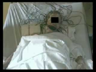
An 86-year-old woman was admitted for a rapid loss of autonomy occurring over one week, as a result of stereotyped, brief, sweeping movements. She was born at term of nonconsanguineous parents. There was no hypertonia at birth and neurodevelopment and growth were normal. She described an exaggerated startle reflex since adolescence, but which had never been accompanied by falls. She jumped every time the telephone rang, a car horn sounded, or if someone knocked on the door, although she never thought that this was abnormal because it caused no functional impairment. Her past medical history was positive for hypertension, coronary disease and peripheral arteriopathy. She had also undergone a hysterectomy and appendectomy. She reported no familial history of exaggerated startle response. At admission, she had been taking clopidogrel, losartan, naftidrofuryl, and molsidomine over a long period. She reported stereotyped short movements one week before hospitalization, induced by unexpected noise. Progressively, abnormal movements occurred for every noise or other unexpected stimuli until she was unable to walk, stand up, or eat. Upon admission, the clinical examination showed proximal jerks that began with blinking followed by facial grimacing, flexion of the head, flexion of the arms toward the face, and extension of the trunk and legs. Startle responses were induced by sudden auditory, visual or tactile stimuli, especially in the form of brief taps on the nose, but also taps applied to other areas of the body. At that point, she stated that the startle could occur in response to her own voice. Neurological examination found generalised hyperreflexia, but plantar responses were flexor. CT brain scan was normal. Brain MRI could not be performed because of jerky movements induced by noises, even under treatment. Video EEG combined with EMG during repeated tapping of the nose demonstrated the excessive startle response and absence of epileptiform activity. The back-averaging study demonstrated the absence of EEG transient activity preceding the EMG activity. Typically, the eyes closed and the face tensed into a grimace. The head flexed forward and the arms took on a defensive posture with abduction of the shoulders, flexion of the elbows, pronation of the forearms and clenching of the fists (see video). Average onset latencies of EMG activity after a nose tap were: 20 ms for orbicularis oculi, 50–60 ms for sternocleidomastoid muscle and 90 ms for deltoid muscle (( figure 1 )). Lack of habituation during repetitive tactile stimulation was demonstrated (( figure 2 )).Treatment with clonazepam (0.2 mg three times a day) reduced the frequencyand intensity of startle, so that she was again able to live alone at home, at the last follow-up of 18 months.
Discussion
The present case illustrates the occurrence of hyperekplexia in the elderly. The recorded events were compatible with the clinical diagnosis of startle reflex. Moreover, neurophysiological investigation confirmed this diagnosis, but showed several features which differed from normal startle: lack of habituation (absence of decrease in both duration and integrated EMG), and prolonged duration of EMG bursts, suggesting the diagnosis of hyperekplexia. On the other hand, it was not possible to distinguish two separate components in the EMG response in orbicularis oculi as, shown by others [6]. The normal startle response consists of a rapid, patterned motor activation in response to a sudden, surprising stimulus. With normal development, the startle response undergoes a considerable modulation by higher limbic and cortical influences so that its intrusion into everyday life is usually minimal [1,7]. The muscular jerk of startle is quick enough to satisfy the definition of myoclonus. The earliest component of the acoustic startle response (the commonest way to assess startle response) is assumed to be generated within the brainstem. The ventral cochlear nucleus projects to the auditory relay neurones within or near the ventral nucleus of lateral lemniscus. This nucleus in turn projects to the pontomedullary reticular formation, regarded as the motor effector area of the startle response [1,6,8]. Onset latencies of EMG activity to unexpected auditory stimulus are: orbicularis oculi, 30–40 ms, masseter and SCM 55–85 ms, biceps brachii 85–100 ms, and quadriceps 100–125 ms [6]. Interestingly, and to the best of our knowledge, no mean latency after the nose tap has been described. EMG burst duration variesfrom 50 to 400 ms, becoming shorter after habituation, and a typical pattern of synchronous activation of antagonist muscles is usually recorded [2,3]. Physiological startle reflexes demonstrate high plasticity [1], with habituation leading to a decreased magnitude of response following repetitive presentation of triggering stimulus; sensitization resulting in increased amplitude or decreased latency of startle response under repeated stimuli (the larger simultaneous effects of habituation often mask it); prepulse inhibition, meaning that the amplitude will be reduced when a brief, nonstartling sensory stimulus precedes a startle auditory stimulus by 30–200 ms; and finally, fear-conditioned potentiation.The term hyperekplexia is often used synonymously with any presumed startle disorder. In the patient reported here, etiological investigations remained negative. Moreover, the lack of any relevant personal and familial past medical history and the absence of any associated neurological involvement were suggested the diagnosis of idiopathic hyperekplexia. The life-long history of excessive startle response reported by the patient favours this putative diagnosis. However, given the limited spatial value of CT scan exploration, we could not exclude a restricted brainstem infarction. A brain MRI would have required deep anaesthesia, which was rejected as potentially harmful because of the patient’s age and the spontaneous favourable outcome.
Hyperekplexia can be an hereditary neurological disorder characterised by continuous generalised stiffness in the first year of life and exaggerated startle reflex, accompanied by temporary generalised stiffness and falls [5,6].
Mutations in the gene encoding the alpha 1 subunit of the glycine receptor (GLRA1) are responsible for the major form [5,6], while a minor form, with only exaggerated startle, could be associated within a single family, without mutation [9]. Sporadic cases do not seem to have a genetic basis and most are symptomatic as a consequence of acute brainstem pathology [10,11,12].
Brainstem disease may be associated with hyperekplexia, as reported in the literature for inflammatory lesions [10], subacute encephalomyelitis with lymphocytic infiltration of the pons and medulla [11,12], brainstem haemorrhages, or Chiari malformation (improvement of hyperekplexia after surgical decompression). Cerebral or diencephalic lesions such as perinatal hypoxic ischaemic encephalopathy and inflammatory, traumatic or vascular thalamic lesion could also induce hyperekplexia [13]. Psychiatric disease, Gilles de la Tourette’s syndrome [14] (during which exaggerated startle response is reported to occur in approximately 5% of patients) and drug intoxication such as maternal cocaine exposure may be associated with startle disease. We are not aware of sporadic cases of exaggerated startle that have occurred so late in life in the absence of any etiology, as with this patient. However, she had a long history of vascular disease, suggesting that a brainstem stroke could not be excluded. She had not exhibited any other symptoms suggestive of brainstem involvement (cranial nerve palsy, drowsiness, cerebellar disturbances, etc.) and she had presented with excessive startle since a young age. We did not look for the GLRA1 mutation since the major form linked to the mutation was not considered a valid possibility, given the absence of other clinical signs and the absence of familial cases. The good and rapid response to low-dose clonazepam suggested a functional disorder rather than an organic lesion.
Late-onset hyperekplexia may be associated with a serious disease that should be extensively investigated, but idiopathic cases can be seen even in elderly patients and constitutes a treatable cause of rapid and dramatic loss of autonomy.