Epileptic Disorders
MENUIctal movement disorders and hypothalamic hamartoma Volume 7, issue 1, March 2005

Auteur(s) :, William Szurhaj, Christine Daems-Monpeurt, Haouaria Sediri, Alain Destée, Philippe Derambure*
Department of Clinical Neurophysiology and Neurology, CHRU
Lille, France
Article reçu le 12 Février 2004, accepté le 6 Novembre 2004
Motor manifestations are a frequent expression of epileptic seizures. They usually consist of clonic, tonic and tonic-clonic movements, dystonia or motor automatisms. To the best of our knowledge, there is no report to date of epileptic choreic/ballistic movements. Such choreic/ballistic movements are classically observed in basal ganglia disorders (subthalamic nucleus or striatum) (Shannon 1990, Albin 1995). We report the case of a woman who presented ictal choreic/ballistic movements. Epilepsy was related to a hypothalamic hamartoma (HH). Since the hypothalamus is not known to be involved in motor command, we discuss the possible pathophysiology of the observed movement disorders in general, and the links between the epileptic discharges and the basal ganglia in particular.Case report
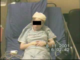
The patient was a 32-year-old, left-handed woman presenting with refractory epilepsy. She was married and had one child. There was no family history of epilepsy. Birth and early developmental milestones had been normal but our patient had experienced some difficulty in following a normal school programme (at the age of 16, she obtained a vocational training certificate in hairdressing). She began suffering from seizures at the age of 12, and these were described as involving a sudden desire to laugh or cry, and associated with anxiety and face flushing. This was followed by abnormal movements of the left side, consisting of tonic hyperextension of the upper or lower left limb (which sometimes caused a fall). The manifestations were brief, lasting about 10-20 seconds, and were first considered to be a psychiatric disorder: epilepsy was only diagnosed when she was 16, after the occurrence of secondarily generalized seizures. The patient had to stop work as a cleaner at the age of 19 because of epilepsy. A CT scan performed when 16 years old was interpreted as being normal. The patient was treated, first with valproate and then with carbamazepine, which resulted in control of the secondary generalization only. At the same time, the episodes of laughing and crying disappeared. At 18 years of age, she was hospitalized in the psychiatry department for behavioural disorders and depression. Gabapentin, topiramate, carbamazepine and lamotrigine were tried without success. Seizures remained frequent during the daytime (between one and 10 per day, more frequent during menstruation), but rare at night. The seizures totally disappeared when she was pregnant but recurred immediately after delivery. Brain MRI revealed a large, sessile, hypothalamic hamartoma of the tuber cinereum ( (figure 1) ). The patient was referred to our unit for exploration of this partial, refractory epilepsy. She received carbamazepine (800 mg/day) and lamotrigine (400 mg/day). The neurological examination, neuropsychological assessments, neuro-ophthalmological and neuro-endocrinal tests were normal. There was no precocious puberty. Long-term video/EEG monitoring was carried out over five days. Interictal EEG showed a well-organized background activity with occasional sharp waves over the central regions, which were activated during sleep ( (figure 2) ). Six brief seizures were recorded, three of which are presented (video). At seizure onset, the patient reported an indefinable feeling within her head, together with a hot flush. She presented polypnea, slight agitation with waddling movements and sometimes a swaying of the trunk. Her eyes were wide open and her face became red. However, the patient did not feel anxious; she remained aware throughout the seizure and was able to push a button to alert nurses, but she was not able to speak. She then reported tension in the left shoulder (and sometimes in the lower limb); agitation then increased, with occasional pedalling movements and other movement manifestations of the left upper limb. The latter were complex (see video recordings) and associated with initial, brisk, proximal, ballistic-like movements, followed by slower, more distal movements, similar to choreic/dystonic movements. Ictal EEG only showed a diffuse depression starting at clinical onset, and lacking paroxysmal abnormalities or rhythmic activity ( (figure 3) ). Interictal single photon emission computed tomography (SPECT) indicated only slightly reduced bilateral, frontal perfusion. Rather than showing hyperperfusion, ictal SPECT revealed a broad zone of hypoperfusion covering the bilateral frontal regions and basal ganglia ( (figure 4) ).We inferred partial, symptomatic epilepsy related to the HH. The patient will soon undergo gamma-knife surgery.
Discussion
Hypotalamic hamartoma is a known cause of epilepsy, and is associated with mental retardation and precocious puberty (Berkovic et al. 1988). The onset of epilepsy is usually marked by gelastic or dacrystic seizures. In our case, the first interesting feature is the observation that gelastic seizures may only be present transiently at the onset of the condition. When we saw the patient, she had been free of gelastic seizures for 15 years. Moreover, she was not mentally retarded and had not presented with signs of precocious puberty. This may well be explained by the sessile form of the hamartoma (Arita et al. 1999). Secondly, ictal motor manifestations are atypical. Vascular causes can be excluded because of the repetitive nature of the manifestations and the absence of vascular lesions on the MRI. Medication cannot produce this type of movement as a side-effect. Psychogenic seizures are unlikely, in light of the video-EEG data, ictal cerebral blood flow changes, the diffuse EEG depression and the absence of psychological disorders. Hence, the choreic/ballistic movements can be interpreted as being of epileptic origin.To the best of our knowledge, such movements have never been described in HH. The pathophysiology of such complex movement disorders remains difficult to explain. It has already been demonstrated that gelastic seizures can originate from within an HH (Munari et al. 1995, Kuzniecky et al. 1997, Kahane et al. 2003), although not all types of seizures can arise from the hamartoma itself (Freeman et al. 2003).
In our case, the gelastic seizures, the interictal EEG and the ictal SPECT results argue in favour of an origin within the HH. The choreic/ballistic movements are more difficult to explain. The hypothalamus has no demonstrated role in the control or execution of movement. Moreover, there is no description of efferent connections to either the cortical sensorimotor areas or the basal ganglia. Hypothalamo-spinal connections do exist but they project to the autonomic nuclei and not to motor neurons. Thus, the involvement of the hypothalamus in movement disorders could be indirect, via other structures. Hemiballismus is most often due to lesions of the subthalamic nucleus. However, in some cases, cortical (Buruma and Lakke 1986) or striatal lesions (Destee et al. 1990) can play a role. In this later case, hemiballismus is often associated with choreic movements (hemichorea-hemiballismus), such as those observed here. Therefore, a prime hypothesis would be the suppression of inhibition of thalamic activity by the pallidum (Shannon 1990, Albin 1995). Moreover, it has already been suggested that dyskinesia-like seizures may involve the basal ganglia (Vercueil and Hirsch 2002). However, even though our patient’s HH was of a considerable volume, it did not extend to these structures, and there are no direct efferents from the hypothalamus to the basal ganglia. On the other hand, we do know that there are many projections from the hypothalamus to the cortex (and to the frontal and temporal lobes in particular): it has been demonstrated that clinical manifestations of HH-related seizures are often due to the involvement of these cortical areas (Berkovic et al. 2003, Andermann et al. 2003). In our case, a number of observations suggest the involvement of the internal part of the frontal lobe (and particularly the supplementary motor area): i) the bilateral, interictal, central, sharp waves; ii) the diffuse depression of ictal EEG; iii) the motor signs and iv) the speech arrest. We know that the SMA is one of the main input structures of the basal ganglia. We therefore speculate that in our case, the ictal discharge starts in the HH, spreads to the internal frontal lobe and then propagates into the basal ganglia network. What happens to the epileptic discharge within the basal ganglia remains unclear. The question of the transmission of epileptic discharges to the basal ganglia remains a subject of debate. Lombroso (1995) has reported recording an epileptic discharge within the caudate nucleus in a girl with paroxysmal kinesigenic choreoathetosis. However, Rektor et al. (2002) recorded 16 seizures using depth electrodes in the putamen, pallidum, and caudate nucleus and did not detect ictal discharge within the basal ganglia (merely an EEG slowing). In our case, ictal SPECT showed hypoperfusion of the basal ganglia rather than hyperperfusion, which is an additional argument against an epileptic discharge within the basal ganglia. The EEG slowing could signify functional disorganization of the basal ganglia network, which would result in abnormal, involuntary movements. We speculate that the epileptic cortical projections produce hyperexcitation of the striatum, which would cause hyperinhibition of the internal globus pallidus and therefore suppression of the inhibition of thalamic activity by the outer pallidum, thus allowing the occurrence of the choreic/ballistic movements (( figure 5 )).