Epileptic Disorders
MENUGAD65 antibody-associated autoimmune epilepsy with unique independent bitemporal-onset ictal asystole Volume 20, issue 3, June 2018

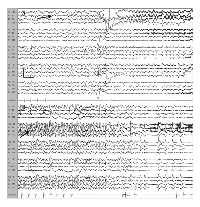
Figure 1
EEG-ECG tracings from a patient with GAD65 antibody-associated autoimmune epilepsy with unique independent bitemporal-onset ictal asystole, showing a left temporal-onset focal seizure discharge with a sentinel sharp wave (not shown), followed by rhythmic left temporal delta activity associated with ictal asystole (A; arrow) and a right temporal-onset focal seizure with ictal asystole (B; arrow).

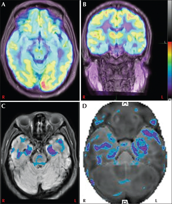
Figure 2
Axial (A) and coronal (B) fused PET MRI showing bitemporal (left>right) hypometabolism. Axial fused PET MRI (C) and surface rendering (D) in the same patient shows significant clustering in the bitemporal lobes (left>right). L: left; R: right.

