Epileptic Disorders
MENUExceptionally long absence status: multifactorial etiology, drug interactions and complications Volume 1, issue 4, Décembre 1999

|
|
||
|
|
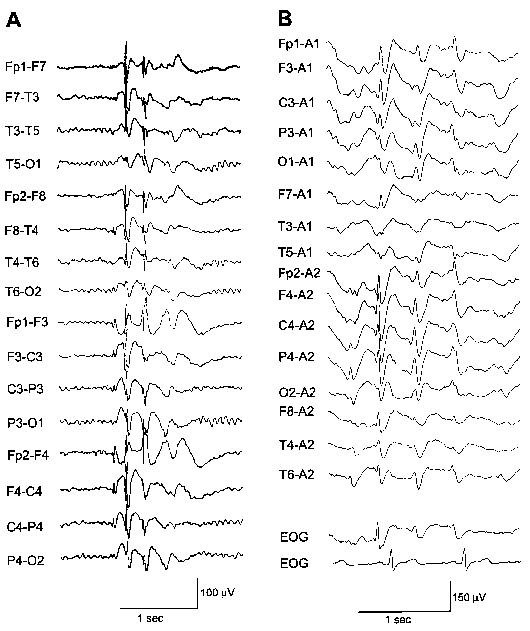
Figure 1. A. Pre-status EEG, "Queen square-parasagittal" montage. Generalized spike and waves at 3 Hz with anterior head regions predominance. Background activity consists of a well-regulated 9 Hz alpha rhythm. B. EEG during status, referential montage to the ipsilateral ear. Generalized slow spike and wave complexes at 2-3 Hz, interspersed with rhythmic high amplitude 2-3 Hz delta waves. |
|
|
|
||

|
|
||
|
|
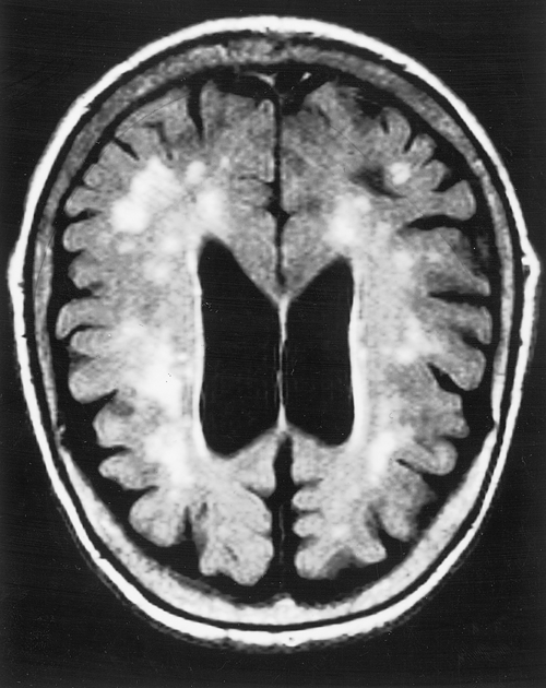
Figure 2. MRI of the head, axial FLAIR sequence showing cortical and subcortical athrophy and hyperintense lesions scattered through the white matter of the centrum semiovale, compatible with ischemic leucoencephalopathy. |
|
|
|
||

