Epileptic Disorders
MENUEncephalopathy with epileptic spasms resolved with corticoids in a 69 year-old patient Volume 2, issue 2, Juin 2000

Epileptic spasms are generalized seizures characterized by a brief axial muscular contraction often occurring in clusters [1]. When associated with hypsarrhythmia and mental deterioration they define West syndrome [2, 3]. Epileptic spasms begin typically before 1 year of age [3-5], although onset in childhood has been reported [4, 5].
We report a 69 year-old man who presented epileptic spasms, with features strinkingly similar to the epileptic spasms recorded in infancy, during the course of encephalopathy of unknown etiology.
Case reports
The patient is a 69 year-old, right-handed, retired truck driver with no particular past personal or familial medical history. He was evaluated the first time for an episode of loss of consciousness. During the few months preceding this event, his wife had noticed his increasing fatigue, throbbing headaches, lack of concern, incoherent conversation especially in the evening, and problems with remembering recent events. Neurological examination was normal but on mental status examination the patient was disoriented in time and place, and had moderate retrograde amnesia for several weeks. He was slow in executing motor commands and showed perseveration on tests of frontal lobe function. His speech was slow but correct, and there was no agnosia. Left temporal sharp waves and two left temporal seizures were recorded on the EEG. Seizures involved sudden loss of consciousness, oro-alimentary automatism and palilalia, followed by clonic movements of the right arm. Moderate hyponatremia (129 mmol/l, N: 135-145), hypo-osmolality between 265 and 285 mmol/kg (N: 285-295 mmol/kg), and a moderate vitamin B1 and B6 deficiency were reported. Carbamazepine, vitamins and aspirin were introduced.
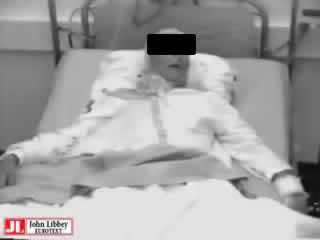
In the following months, episodes of confusion occurred and were attributed to worsened hyponatremia (124 mmol/l). Clinical examination revealed a bilateral Babinski sign, short and long term memory deficit, constructive apraxia and temporo-spatial disorientation. Water intake restriction and introduction of phenytoin instead of carbamazepine were proposed. Vigilance status worsened despite control of serum sodium and 6 months after the first hospitalization the patient became comatose (Glasgow 4) with episodes of Cheyne-Stokes respiration. EEG background was permanently slow (0.5-4 Hz) with abundant triphasic waves. The EEG amplitude ranged between 60 and100 muV. No focal seizures were recorded during this period but frequent episodes of spasms recurred despite treatment with valproate, phenobarbital, phenytoin, and clonazepan. Spasms consisted of sudden flexion of the neck and all four extremities, with adduction of the arms. They were symmetric or slightly asynchronous, starting on the left as assessed by polygraphic study (figure 1). Wide opening of the eyes and occasional moaning were observed. Spasms were sometimes repeated after 2 seconds and recurred every 0.5-3 minutes in long clusters. Ictal EEG (figure 1) showed diffuse high amplitude slow wave and fast activity lasting 0.5-1.5 s preceded and followed by a diffuse attenuation of the EEG for few seconds.
MRI at 1, 6 and 11 months of follow-up showed abnormal bilateral hypersignals (L > R) in the white matter in T2-weighted images. Ventricle size and morphology were normal. There was moderate, predominantly supratentorial, cortical atrophy. Echo Doppler study of the carotid arteries only showed mild atheromatosis of the carotid bifurcation. Cardiac echography was normal. Cerebral angiography showed signs of neither atherosclerosis nor vasculitis. Cerebrospinal fluid (CSF) examination was normal, except for a relative increase of plasmocytes (6%). Electromyography showed no signs of myopathy. Biological evaluation (sedimentation rate, C reactive-protein, antinuclear and antithyroid antibodies, VDRL, vitamin B12, cortisol, conversion enzyme inhibitor, lactate, pyruvate, porphyrins, extensive viral serologies in blood and CSF, CSF two dimensional electrophoresis), thoracic and abdominal CT-scans, gastrographin abdominal X-radiographs were normal. Ictal single photon computed tomography (SPECT) of regional cerebral blood flow performed with Tc99m-HMPAO during the ictal phase, showed bilateral hyperperfusion of the head of the caudate nuclei and of the right pallidum as compared to the interictal SPECT (figure 2). Positron emission tomography with F18-fluorodeoxyglucose (FDG-PET) performed during the comatose state with frequent spasms revealed hypermetabolism in the caudate nuclei (figure 3).
After 10 days of coma, methylprednisolone (500 mg/day i.v. for 5days) followed by prednisolone (60 mg/day) were introduced. Vigilance was dramatically improved and the spasms disappeared. Neuropsychological examination progressively returned to normal except for long term memory disturbances and a discrete constructional apraxia. Temporal lobe seizures recurred but were easily controlled by VPA. After a few weeks, oral steroids were replaced by azathioprine (100 mg a day) which is still prescribed (2 years follow-up). As immunosuppressive drug withdrawal trials induced important cognitive disorders and increased frequency of partial seizures, treatment was quickly reintroduced.
Discussion
The epileptic spasms recorded in our patient present striking similarities with the seizures typically described in West syndrome during infancy. The semiology of the brief, sudden, and repetitive episode of flexion closely resembled that of the flexor spasms recorded during infancy. Ictal electroencephalographic findings characterized by high amplitude slow wave intermixed with fast rhythmic activity during the spasm were equally comparable. There was associated major cognitive impairment. Furthermore, precession by partial seizures is described in symptomatic West syndrome [3, 4, 6]. As often described during infancy, focal features disappeared in our patient with worsening of the general condition, only to reappear after the comatose period, with spasms suggesting a secondary generalization of partial seizures [3, 6]. The poor efficacy of conventional antiepileptic drugs and the dramatic improvement after corticoids are observed in infantile spasms [7, 8].
Recently, based mostly on PET studies, the hypothesis of cortical-subcortical interaction has been proposed to explain the physiopathogenesis of epileptic spasms [3, 9]. In our patient, both PET study and ictal SPECT showed basal ganglia activation as described in infantile spasms studies [9, 10].
The etiology of the subacute encephalopathy remains unknown. Based on the clinical and radiological picture, and the response to corticosteroids and immunosuppressive drugs, a vasculitic origin was hypothesized. However, thorough investigation did not result in a positive diagnosis and the family refused brain biopsy.
The recognition of the striking similarities between the seizures recorded in our patient and the epileptic spasms of West syndrome has important therapeutic consequences. Corticoids should be used rapidly in adult patients with similar electroclinical features after failure of conventional antiepileptic drugs.
Received May 16, 2000 / Accepted May 30, 2000