Epileptic Disorders
MENUElectrical status epilepticus “invisible” to surface EEG in late-onset Rasmussen encephalitis Volume 10, issue 3, September 2008

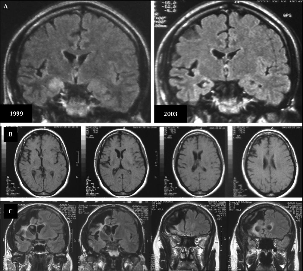
Figure 1 MRI images (A) coronal T2-weighted FLAIR images show a progressive, right hemispheric atrophy predominantly involving the temporal lobe (a hyperintensity in the right hippocampal region is also evident in the first scan), and the perisylvian regions; (B) axial T1-weighted images document the extensive involvement of frontal lobe and perysilvian region in pre-surgical evaluation; (C) post-surgical MRI scan: T2-weighted FLAIR images show a wide right fronto-dorsal corticectomy.

Figure 2 A) Continuous periodic epileptiform abnormalities configuring an electrical status epilepticus located in the right fronto-dorsal regions; panels show an enlargement of the EEG tracings: electrical status, which is well documented by the subdural grid (upper panel), is not detected by the corresponding surface recording (lower panel), which only shows slow theta-delta monomorphic activity. B) a seizure triggered by the aforementioned epileptic electrical activity, arising from the same fronto-dorsal regions and spreading to the homolateral temporal lobe and interemispheric structures; panel shows an enlargement of the corresponding EEG tracings. C) Schematic illustration of the subdural grid location: red points indicate electrodes corresponding to the subcontinuous ictal activity.