Epileptic Disorders
MENUConcept of epilepsy surgery and presurgical evaluation Volume 17, issue 1, March 2015
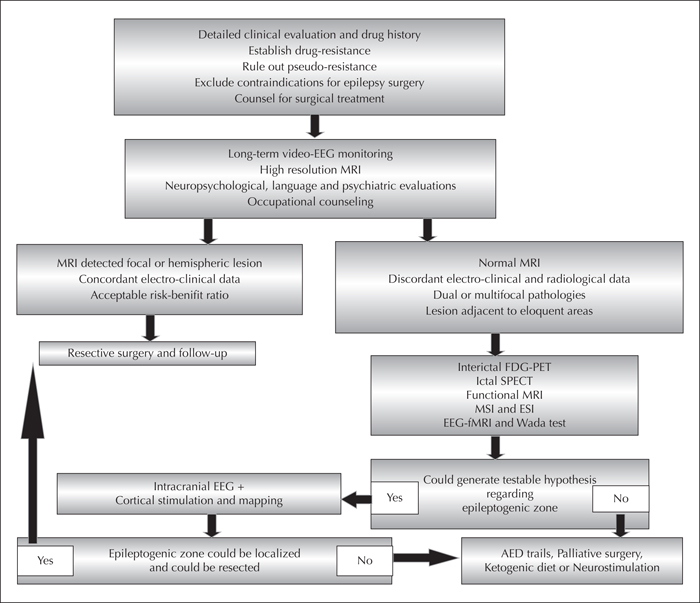
Figure 1
Flow chart depicting the usual process of presurgical evaluation and selection for surgery (reproduced with permission from Rathore et al., 2014).
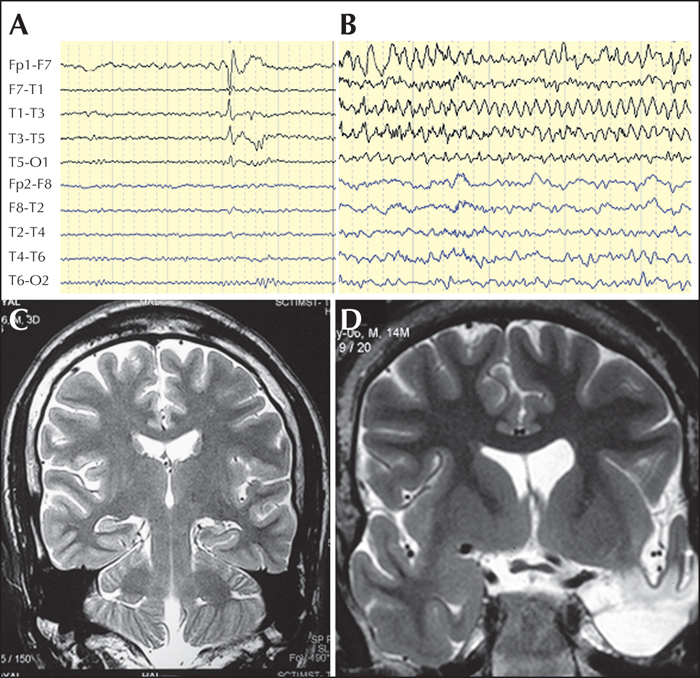
Figure 2
The EEG-MRI data from one of the operated patients: (A) EEG showing left temporal interictal epileptiform discharges; (B) 7-Hz-rhythmic theta activity over the left temporal region; (C) left hippocampal atrophy and increased signal on T2-weighted MRI sequence; and (D) postoperative MRI showing complete resection of mesial temporal structures. The patient was seizure-free for over five years following left anterior temporal lobectomy with amygdalo-hippocampectomy.
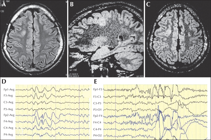
Figure 3
Presurgical non-invasive data from a patient with drug-resistant frontal lobe epilepsy: (A) axial FLAIR image showing thickened cortex and signal changes over the right frontal region; (B) saggital 3-D FLAIR image showing extension of the lesion to periventricular region (transmantle dysplasia); (C) functional MRI showing left hand motor function immediately posterior to the lesion; (D) interictal recording showing right frontal spike discharges; and (E) ictal EEG recording showing right frontal beta activity at the onset of a complex partial seizure. The patient did not develop any neurological deficit following lesion resection and has been seizure-free for the last five years.
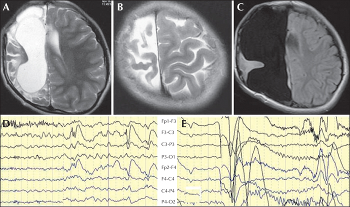
Figure 4
Presurgical data from a patient with drug-resistant seizures and minimal weakness of the left hand: (A, B) T2-weighted axial MRI sequences showing extensive encephalomalacia over the right hemisphere with relatively preserved motor cortex; (C) postoperative axial FLAIR image following hemispherectomy with preservation of motor cortex; (D) interictal EEG showing relative suppression of background activity over the right hemisphere with bifrontal spike-and-wave discharges which appeared more prominent over the left frontal region; and (E) ictal EEG showing diffuse attenuation of background activity for one second, followed by appearance of diffuse 14-Hz activity. The patient was seizure-free for two years without any additional motor deficits.
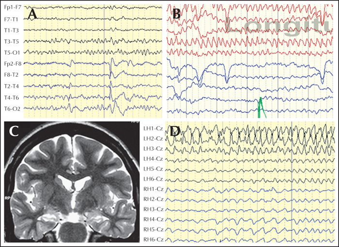
Figure 5
Invasive evaluation in temporal lobe epilepsy: (A) interictal scalp EEG showing right temporal spike discharges; (B) ictal scalp EEG showing left temporal 6-Hz rhythmic theta activity during a complex partial seizure; (C) T2-weighted coronal MRI showing bilateral mesial temporal sclerosis, slightly more pronounced on the left side; and (D) invasive EEG monitoring with bilateral hippocampal depth electrodes (inserted orthogonally in postero-anterior direction; LH1: left hippocampal contact 1) showed origin of all the 11 recorded seizures form the left hippocampus. Neuropsychological evaluation revealed predominant verbal memory impairment. The patient has been seizure-free for the last four years and did not have any significant worsening of memory.

