Epileptic Disorders
MENUAbdominal epilepsia partialis continua in neurocysticercosis Volume 21, issue 3, June 2019


Case study
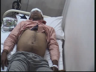
Epilepsia partialis continua (EPC) is characterised by continuous involuntary jerky movements restricted to a part of the body with preserved awareness. EPC of abdominal muscles is a rare entity with variable clinical localization and aetiology. A 25-year-old man presented with sudden onset of arrhythmic focal myoclonic movements exclusively involving the abdominal muscles on the right side, lasting between 20 minutes and an hour. He had no prior history of seizures and had a normal birth and developmental history. He had no known medical comorbidities or addictions. The neurological examination was normal except for focal myoclonus over the abdomen seen continuously with no involvement of the face, arms or lower limbs (video 1). Contrast-enhanced brain MRI revealed a ring-enhancing lesion, suggestive of cysticercal granuloma over the left precentral gyrus (figure 1A, B, C). EEG did not show focal abnormalities, either during the events or in the interictal period (figure 2). Serological testing for neurocysticercosis was offered to the patient but declined due to financial constraints. The patient fulfilled the revised diagnostic criteria for definitive diagnosis of neurocysticercosis (Del Brutto et al., 2017). He was initially started on oxcarbazepine and subsequently AEDs were up titrated to achieve seizure control. Episodes of EPC were controlled with difficulty using 600 mg oxcarbazepine, 200 mg lacosamide, and 2,000 mg levetiracetam. He received antiparasitic therapy with albendazole (15 mg/kg for two weeks) and oral dexamethasone (0.1 mg/kg) for two weeks which was then tapered. Follow-up EEG was performed at one month and three months which showed slowing over the left frontal region. The patient continues to have rare short-lasting episodes of abdominal EPC and focal motor seizures at a frequency of 1-2 episodes per month.
Discussion
EPC was first defined by Kozhenikov in 1894, but it was Thomas et al. (1977) who first defined EPC based on its phenomenology. According to this definition, episodes of EPC are spontaneous, regular or irregular, clonic muscular twitching, affecting a limited part of the body, sometimes aggravated by action or sensory stimuli, occurring for a minimum of one hour and recurring at intervals of no more than ten seconds. The ILAE task force report on status epilepticus considers EPC as a subclass of focal motor status although sensory manifestations are known to occur (Trinka et al., 2015). Our patient had events lasting ≥one hour which fulfils the criteria for EPC in addition to shorter events which qualify as abdominal focal motor seizures.
The pathophysiology of EPC is still unclear. A single mechanism is inadequate to explain the complex manifestations including stimulus sensitivity, however, a combination of epileptogenic motor cortex coupled with abnormal excitation of cortico-subcortical loops is likely to be responsible for producing EPC (Guerrini, 2009). Episodes are commonly distributed over the face and distal upper and lower limbs (Thomas et al., 1977; Sinha and Satishchandra, 2007; Mameniskiene et al., 2011). Abdominal EPC is a rare manifestation and only 12 cases have been reported in the literature to date (table 1) (Matsuo, 1984; Rosenbaum and Rowan, 1990; Chalk et al., 1991; Biraben and Chauvel, 1997; Fernández-Torre et al., 2004; Lim et al., 2004; Dafotakis et al., 2006; Tezer et al., 2008; Ribeiro et al., 2015). The largest series of patients with EPC -66 patients from India (Sinha and Satishchandra, 2007), 51 paediatric patients (Kravljanac et al., 2013), and a European survey of 65 patients with non-stroke and non-Rasmussen's EPC (Mameniskiene et al., 2011),make no separate mention of abdominal myoclonus , although six patients had hemibody involvement in the series by Mameniskiene et al. The limited representation of abdomen over motor homunculus may account for the rarity of this phenomenon (Catani, 2017). It is not a mere coincidence that EPC is more often seen over body parts with proportionately larger cortical representation over the sensory and motor homunculus. Abdominal EPC may occur in isolation or may involve other body parts, as seen in about half of patients in the literature. The lower limbs are most commonly involved, followed by the face, upper limbs, and shoulders. The overlapping representations of body parts, particularly the abdomen and the limbs over the superior aspect of the sensory and motor homunculus, may account for this co-existence (Catani, 2017).
Abnormal MRI is noted in a majority of patients with abdominal EPC and the localisation is commonly to parietal or frontal lobes. MRI in our patient showed a granuloma in the left pre-central gyrus which correlated fairly well with the representation of the abdomen over the motor homunculus. Though this may not be true in all cases, a contralateral parasagittal or medial location is evident in most cases. These observations point to an epileptogenic network which is highly localised and closely involves the peri-Rolandic cortex and subcortex.
The aetiology of abdominal EPC is more variable compared to EPC involving other distributions, of which Rasmussen's encephalitis is a major cause. While the commonest aetiology is vascular, other causes include FCD, aspergilloma, and tumours. This is the first report of inflammatory granuloma due to neurocysticercosis causing abdominal EPC. In a previous large series of EPC from India by Sinha et al., granulomas accounted for 7% although there was no abdominal EPC reported in this study (Sinha and Satishchandra, 2007).
EEG was normal in our patient, which is reported in about a fifth of patients. This is maybe because the cortical activity is very well localised or because of the unfavourable angle of orientation of the dipole in relation to the recording electrodes (Mameniškienė and Wolf, 2017).
Based on its evolution, EPC lends itself to categorisation into one of the following subtypes:
- –(1) EPC as a solitary event
- –(2) Chronic repetitive non-progressive EPC
- –(3) Chronic persistent non-progressive EPC
- –(4) Chronic progressive EPC
Our patient had an evolution resembling chronic repetitive non-progressive type. The prognosis of abdominal EPC depends on underlying aetiology and patients with a vascular aetiology generally have short-lived EPC with good response to AEDs. Our patient, at six months of follow-up, still has rare intermittent episodes of EPC despite being on three AEDs, pointing towards a slightly more protracted course for inflammatory granulomas.
To conclude, abdominal EPC is a rare manifestation, usually associated with brain lesions and varied aetiology. Inflammatory granulomas are a rare cause of abdominal EPC and may not respond readily to AEDs.
Acknowledgements and disclosures
We would like to thank Dr. Sudhir C Joseph, Director of St Stephen's Hospital.
None of the authors have any conflict of interest to declare.