European Journal of Dermatology
MENUThe risk of glaucoma associated with phacomatosis cesioflammea and phacomatosis cesioflammeo-marmorata Volume 32, issue 5, September-October 2022

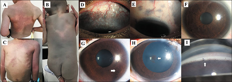
Figure 1.
Segmental dermal melanocytosis and ocular hyperpigmentation in patients with phacomatosis pigmentovascularis. A-C) Extensive grey macules in a “cape” distribution and “bathing trunk” distribution, overlapping with naevus flammeus. D) Episcleral hyperpigmentation presents as diffuse mass-type in a patient with glaucoma and (E) scattered plaque-type in a patient without glaucoma. F) Diffuse iris heterochromia with iris mammillation. G) Obvious hyperpigmentation is detected on the pericornea (white arrow) with a reduced number of iris crypts. H) Punctate hyperpigmentation on the lens (white arrow) and iris heterochromia are shown in a patient treated with 1% tropicamide for mydriasis. I) Gonioscopy reveals increased pigmentation of the anterior chamber angle.

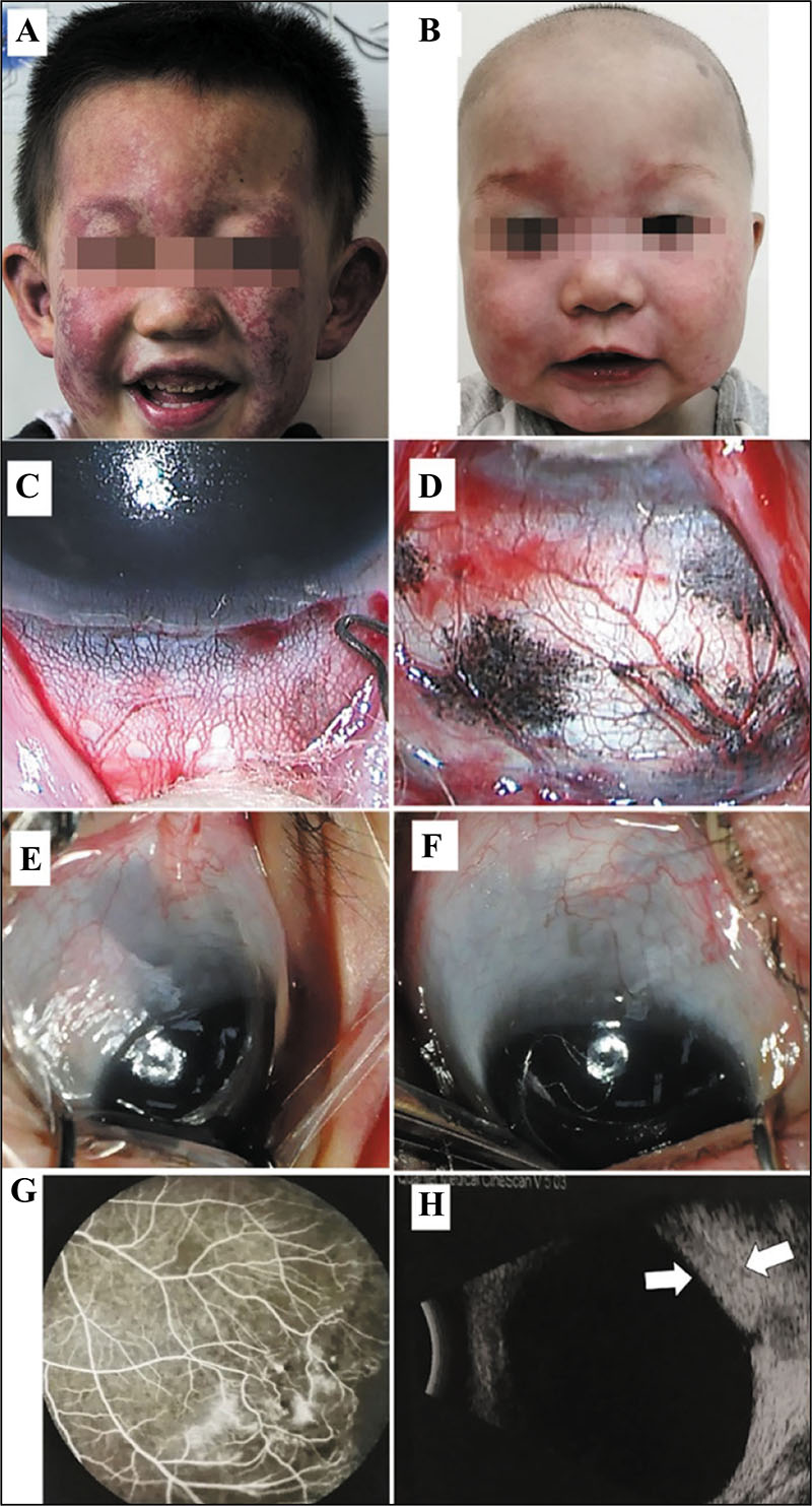
Figure 2.
Capillary naevus and ocular vascular malformation in patients with phacomatosis pigmentovascularis. Facial naevus flammeus is darker in a patient with glaucoma (A) than a patient without glaucoma (B). C, D) Dilated episcleral vessels with increased density present a reticulate appearance with episcleral melanosis. E, F) Dark and total perilimbal episcleral hyperpigmentation are shown in the eyes. G) Fundus fluorescein angiography shows peripheral retinal vascular leakage, venous shunts and tortuous capillaries. H) Choroidal haemangioma (white arrow) demonstrated by B-scan ultrasonography.

