Epileptic Disorders
MENULate adult-onset epilepsy in a patient with hemimegalencephaly Volume 21, issue 2, April 2019

Figure 1
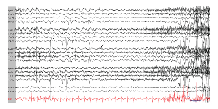
Ictal EEG recording of a developmentally-normal woman who had a first ever epileptic seizure at 55 years old (longitudinal bipolar montage). Rhythmic left frontal activity (arrow) is seen preceding clinical onset. During this event, she had speech reversion to Portuguese, followed by bilateral tonic-clonic activity. All EEG electrodes were placed using the international 10-20 system for electrode placement. Low frequency filter: 1 Hz; high frequency filter: 70 Hz; notch off; sensitivity: 7 μV/mm; time base: 30 mm/s; sampling rate: 500 Hz.

Figure 2
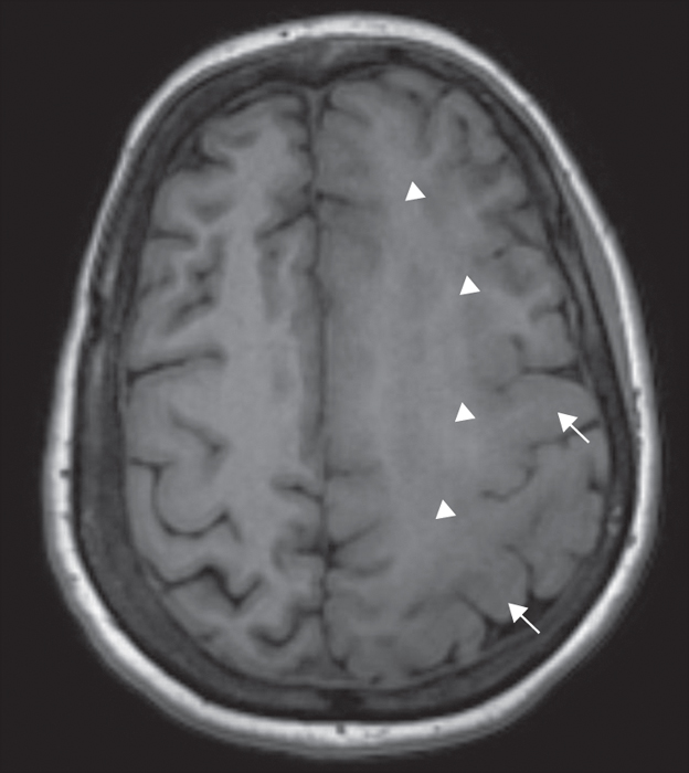
Axial T1-weighted MR images showing left-sided cortical thickening (arrows), deep white matter heterotopias, and abnormal white matter signal (arrowheads), consistent with hemimegalencephaly.

