Epileptic Disorders
MENUKCTD7-related progressive myoclonus epilepsy Volume 18, supplement 2, September 2016

Figure 1
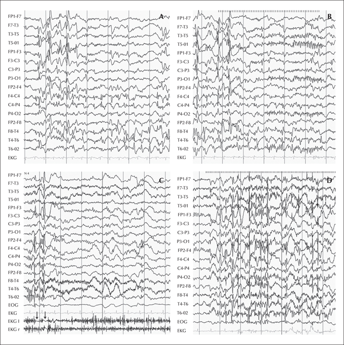
The EEG of Patients 1 and 2 from the original report by Van Bogaert et al. (2007). (A) EEG of Patient 1 at the age of 5 years, awake and at rest, showing slow dysrythmia and multifocal high-amplitude epileptiform discharges. (B) Intermittent light stimulation (ILS) at 12 Hz inducing occipital spikes in addition to the spike-wave complexes still present at rest. (C) EEG of Patient 2 at the age of 14 years, awake with arms maintained in flexion and forearms in extension. On EMG, performed with surface electrodes placed over the left (l) and right (r) deltoid muscles, two negative myocloni appear as flattening of the EMG discharge (arrows), the first one on the left side and the second on both sides. Myoclonus was preceded by right-sided spike-wave discharges, followed by generalized spike-wave discharges. (D) ILS, at 15 Hz, induced generalized spike-waves, together with clinical bilateral myoclonus; these discharges were followed by rhythmic occipital spikes diffusing into a secondary generalized tonic-clonic seizure.

