Sang Thrombose Vaisseaux
MENUPorcelain heart Volume 29, issue 5, September-October 2017

Authors

Hôpital privé d’Antony, Pôle cardiovasculaire interventionnel, 25, rue de la Providence, 92160 Antony, France
* Tirés à part
- Key words: myocardial calcification, computed tomography, congestive heart failure
- DOI : 10.1684/stv.2017.0986
- Page(s) : 203-6
- Published in: 2017
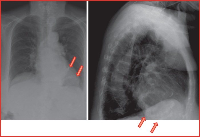
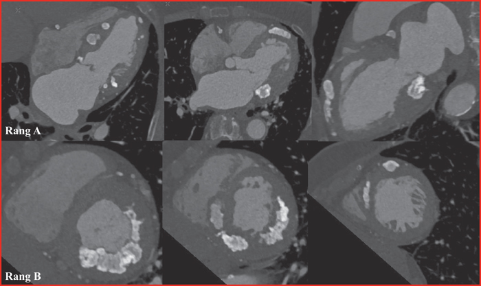
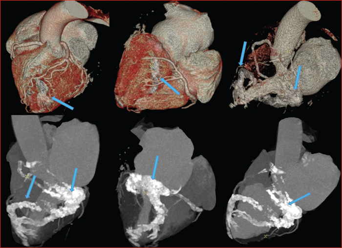
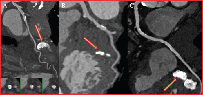
We present a case of massive idiopathic myocardial calcification of the left ventricle in an 81-year-old female who presented with symptoms of congestive heart failure and atrial fibrillation. Massive myocardial calcification is rare and can be classified in metastatic (involving a disturbance in calcium-phosphate metabolism), dystrophic (sequelae of local tissue damage of various origin) or idiopathic. Myocardial calcifications can be detected in radiographs, echocardiography, magnetic resonance imaging but computed tomography is the optimal imaging modality.