Epileptic Disorders
MENUThe neural networks underlying the illusion of time dilation: case study and literature survey Volume 24, issue 3, June 2022

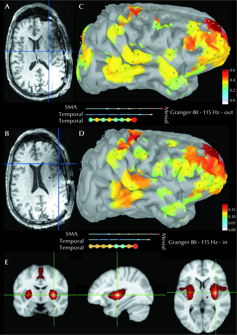
Figure 1.
A, B contacts correlating with time dilation at 4 mA, and 9-11 mA respectively. Axial MRI cuts are oriented in neurological orientation (right is right). Granger causality of the site shown in (A) of highfrequency oscillations 80-115 Hz outflow (C), and inflow (D) in the awake resting state based on icEEG interpolated from electrode space onto the cortex. (E) fMRI connectivity of site correlating with time dilation in radiological orientation.

