Epileptic Disorders
MENUPositron emission tomography in epileptogenic hypothalamic hamartomas Volume 5, issue 4, December 2003

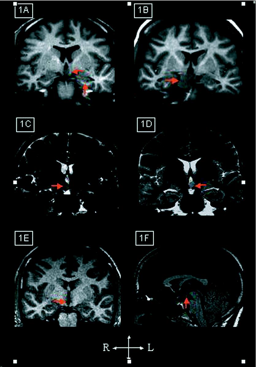
Figure 1. MRI findings.
A) Patient No 1 showing a left-sided,
intra-hypothalamic hamartoma associated with a subtle hippocampal
asymmetry of uncertain significance; B) Patient
No 2 showing a large, right-sided extra-hypothalamic
hamartoma; C) Patient No 3 showing a right-sided,
intra-hypothalamic hamartoma; D) Patient
No 4 showing a left-sided, intra-hypothalamic hamartoma;
E) and F) Patient No 5 showing a
right-sided, intra-hypothalamic hamartoma on both coronal and
sagittal T1 sequences.
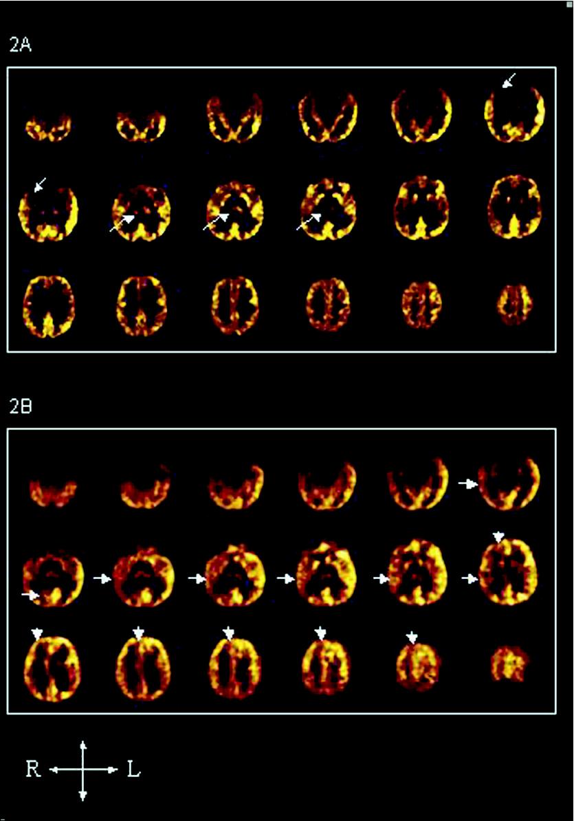
Figure 2. FDG-PET.
A) Patient No 2 demonstrated right thalamic and,
to a lesser extent, right temporo-polar hypometabolism. B)
Patient No 3 showed an extensive, right hemispheric
hypometabolism, involving the lateral temporal cortex, the
orbito-frontal and fronto-polar cortex, the peri-sylvian region,
and the mesial and lateral occipital lobe.
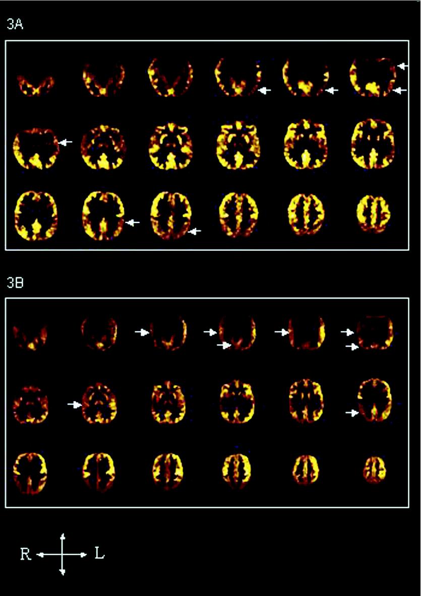
Figure 3. FDG-PET. A) Patient No 4 demonstrated left, lateral, temporo-occipital hypometabolism, but also left parietal abnormalities; B) Patient No 5 showed an extensive, right hemispheric hypometabolism, involving the lateral temporal cortex, the TPO junction, and the inferior parietal lobule.