Epileptic Disorders
MENUPeri-ictal headache due to epileptiform activity in a disconnected hemisphere Volume 16, issue 2, June 2014

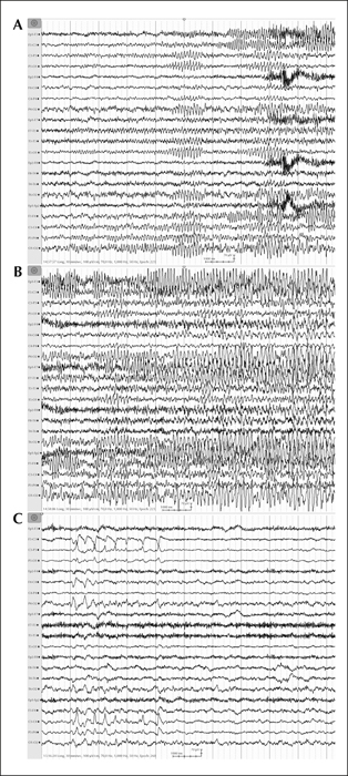
Figure 1
Interictal and ictal EEG recordings at the time of headache attacks.
(A) Seizure onset (from the left posterior quadrant on scalp EEG). (B) Evolving ictal EEG pattern during the headache attack showing the spread of ictal activity. (C) Interictal abnormality with flat non-organised background activity over the left hemisphere and central left/midline spikes.
Longitudinal bipolar montage; amplitude: 150 uV/cm; speed: 10 mm/s.

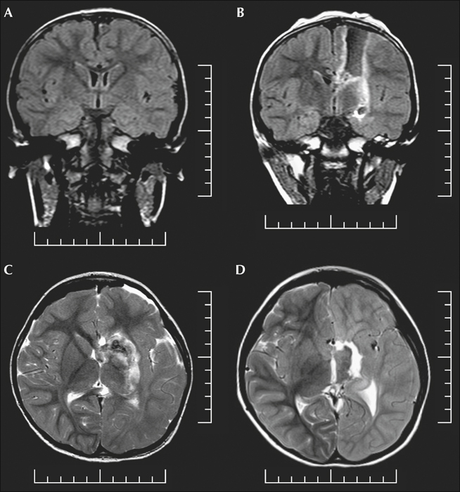
Figure 2
Brain MRI of the patient.
(A) Presurgical coronal FLAIR sequence consistent with a diagnosis of left hemispheric cortical dysplasia. (B and C) Immediate postsurgical coronal FLAIR and axial T2w sequences showing complete disconnection of the left hemisphere. (D) Axial T2w sequence performed at the time of headache attacks revealing enhanced signal changes of the grey and white matter of the left hemisphere.

