Epileptic Disorders
MENUKinesigenic reflex epilepsy associated with a glioma in the lateral peri-rolandic region Volume 11, issue 1, March 2009

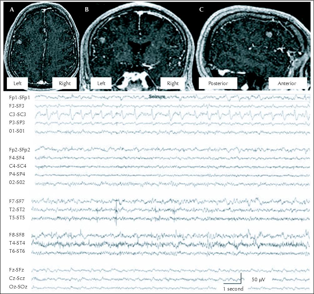
Figure 1 MRI T1-weighted SPGR (spoiled gradient echo) sequences co-localizing the mass lesion with the EEG epileptic network. A) Axial. B) Coronal. C) Sagital. MRI shows a grade III astrocytoma in the left peri-rolandic cortex. EEG is in the Laplacian montage. Sensitivity: 7.0 μV/mm. Paper speed: 30 mm/second. High frequency filter: 70 Hz. Low frequency filter: 1 Hz. 60-Hz filter: off. Epileptiform discharge co-localizes with the mass lesion under the C3 electrode.