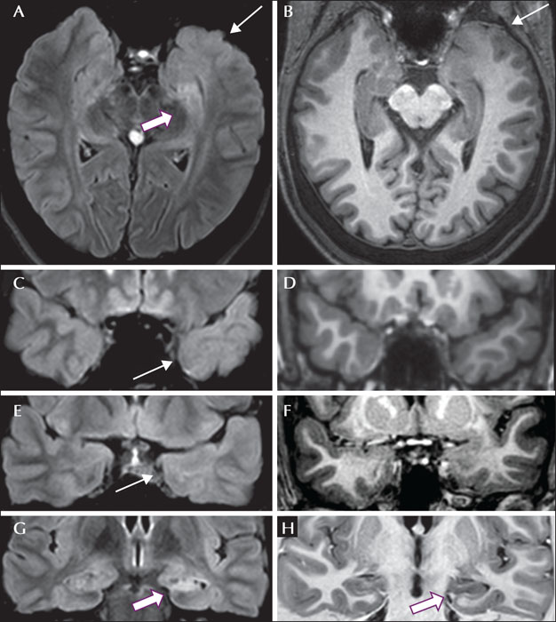
Left hippocampal sclerosis and ipsilateral temporopolar blurring. (A) Axial FLAIR showing hyperintense signal in the hippocampal head (arrow) and ipsilateral temporal pole atrophy with blurring of the transition between gray matter and white matter and hyperintense signal in the white matter (white arrow), also seen on coronal FLAIR images in (C) and (E). (B) Axial T1-weighted image shows discreet changes in the white matter signal with preserved cortical thickness in the temporal pole, also demonstrated on coronal T1-weighted images in (D) and (F). (G) Coronal FLAIR showing left hippocampal atrophy, flattening, and hyperintense signal (arrow). (H) Coronal T1-weighted inversion recovery showing hypointense signal and loss of normal internal hippocampal structure (arrow).
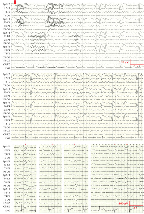
EEG findings. The top panel shows the onset of one of the patient's habitual seizures recorded during video-EEG monitoring (red arrow) on longitudinal bipolar montage. Note the muscle artifacts followed by a rhythmic theta-like activity in the left anterior temporal lobe, maximal at F7-T3 (equipotential) which becomes more evident towards the end of the seizure (middle panel). The lower panel shows samples of interictal slow sharp waves with phase reversals at F7 or equipotential between F7-T3. Different montages also showed maximal involvement at the T1 electrode (not shown here). LFF: 0.3 Hz and HFF: 70 Hz.
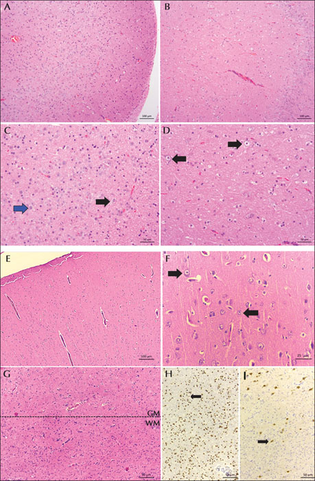
Microscopic features of the hippocampal sectors and the anterior temporal lobe. (A-D) Hippocampal sectors CA1 (A, C) and CA4 (B, D) showing neuronal loss and gliosis. C) Higher magnification of the region shown in (A); the black arrow indicates a remaining pyramidal neuron, and the blue arrow indicates one of the corpora amylacea in the gliotic background. (D) Higher magnification of the region shown in (B) (dentate gyrus on the right); the black arrows indicate remaining neurons. (E-I) Microscopic features of the temporal pole; no histopathological changes compatible with focal cortical dysplasia were evident. (E) Low-power magnification highlighting the preserved architecture of the cortex. (F) Higher magnification of the region shown in (E); the neurons (black arrows) show normal cytologic findings (size, shape, and orientation). (G) Blurring of the gray (GM) and white matter (WM) boundary; the dotted line shows the approximate cortical-subcortical transition. Histopathological analyses showed only an increase in oligodendrocytes (mainly in the WM) and a few heterotopic neurons. (H, I) Immunohistochemical markers for the population of cells in the white matter in (G). (H) Olig-2-positive oligodendrocytes (arrow). I) NeuN-positive neurons (arrow). (A-G) H&E stain; (H, I) peroxidase. Scale bars = 100 mm (A, B and E); 50 mm (C, D, G, H and I); 25 mm (F).
1 Department of Neurology, University of Campinas – UNICAMP, Campinas, SP, Brazil
2 Hotchkiss Brain Institute, Cumming School of Medicine, University of Calgary, Canada
3 Neuroimaging of Epilepsy Laboratory, McConnell Brain Imaging Centre and Montreal Neurological Institute and Hospital, McGill University, Montreal, Canada
4 Multimodal Imaging and Connectome Analysis lab, McConnell Brain Imaging Centre and Montreal Neurological Institute, McGill University, Montreal, Canada
5 Department of Bioengineering, University of Pennsylvania, Philadelphia, USA and Department of Clinical and Experimental Epilepsy, UCL Queen Square Institute of Neurology, University College London, London, UK
6 Institute of Neurobiology, Universidad Nacional Autonoma de Mexico, Queretaro 76230, Mexico
8 The Florey Institute of Neuroscience and Mental Health and The University of Melbourne, Australia
9 Vanderbilt University Institute of Imaging Science, Vanderbilt University Medical Center, Nashville, USA
10 Department of Neurosurgery, University Hospital Erlangen, Germany and Department of Neurosurgery, University Hospital Halle (Saale), Germany
11 Neurology Unit, University of Modena and Reggio Emilia, Modena, Italy
12 Epilepsy Center, Cleveland Clinic, Cleveland, USA
13 Department of Neurology, Second Affiliated Hospital, School of Medicine, Zhejiang University, China
14 Department of Pathology, University of Campinas – UNICAMP, Campinas, SP, Brazil
* Correspondence: Fernando Cendes
Department of Neurology,
Universidade Estadual de Campinas - UNICAMP,
Rua Vital Brasil, 251,
Cidade Universitária Zeferino Vaz,
Campinas, SP. CEP-13083-888, Brazil
We present an illustrative case to address anterior temporal lobe atrophy with poor delineation of the temporopolar gray-white matter interface based on T2-weighted and fluid-attenuated inversion recovery (FLAIR) images in patients with temporal lobe epilepsy associated with hippocampal sclerosis (TLE-HS). A 52-year-old woman with pharmacoresistant seizures since the age of six months underwent a previous MRI scan using a suboptimal protocol which was reported as unremarkable. MRI performed according to an epilepsy protocol showed classic signs of left HS and ipsilateral temporal polar atrophy with blurring of the gray-white matter boundary on FLAIR images. She underwent a left amygdalohippocampectomy and anterior temporal resection and remains seizure-free after 24 months. Histopathological analyses showed HS and no signs of focal cortical dysplasia (FCD). Blurring and atrophy of the ipsilateral temporal pole are common in TLE-HS and often misinterpreted as FCD. This relates to delayed myelination in patients with seizures before the age of two, is more pronounced on FLAIR sequences, and gives a false impression of cortical thickening. However, the T1-weighted images show a relatively well-demarcated cortical-subcortical transition and normal cortical thickness. By contrast, the cortical thickening in FCD is observed on both T1-weighted and FLAIR images. Since FCD also occurs in temporal lobe regions, it is important to differentiate the extra-hippocampal MRI abnormalities in TLE-HS from those likely to be FCD. This case highlights the importance of evaluation based on detailed imaging, which should always be conducted considering the EEG, seizure semiology, and other clinical information.

 This work is licensed under a
Creative Commons Attribution-ShareAlike 3.0 International License.
This work is licensed under a
Creative Commons Attribution-ShareAlike 3.0 International License.