Epileptic Disorders
MENUGelastic seizures: not always hypothalamic hamartoma Volume 9, issue 4, December 2007
Auteur(s) : Christina S Cheung, Andrew G Parrent, Jorge G Burneo
Epilepsy Programme, Department of Clinical Neurological
Sciences, University of Western Ontario, London, Canada
Article reçu le 10 Juillet 2007, accepté le 23 Septembre 2007
Gelastic (laughing) epilepsy has been thought to represent less than 0.5% of all other types of epilepsy (Chen and Forster 1973). Its most common cause is hypothalamic hamartoma, and gelastic seizures are thought to be one of the hallmark features of this condition (Arzimanoglou et al. 2003, Anderman et al. 2003, Kerrigan et al. 2005). However, it has been postulated that they may originate from neural activity in the cingulate gyrus and temporal region as well as the frontal lobe (Loiseau et al. 1971, Arroyo et al. 1993, McConachie and King 1997). Often, MR imaging may not detect a lesion and such cases are deemed cryptogenic (Arroyo et al. 1993, Iannetti et al. 1997, Garcia et al. 2000). There has been study of subtle findings on MRI, associated with cortical dysgenesis and abnormalities of cortical organization that should trigger closer evaluation (Bronen et al. 2000). Ictal EEG can demonstrate various patterns in gelastic seizures of frontal lobe origin, and SPECT has demonstrated hypoperfusion post-ictally, particularly in cases of focal cortical dysplasia (Iannetti et al. 1997, Sasaki et al. 2000).Frontal lobe epilepsy manifests clinically more typically as motor symptoms, which is a useful distinguishing characteristic of frontal versus temporal lobe epilepsy (Geier et al. 1977). Much discussion has surrounded the affective component of gelastic seizures, with feelings of mirth associated with seizures from the temporal lobe. Mirth or pleasant sensations are not usually present in cases of frontal origin (Arroyo et al. 1993). It is worth noting however, that temporal lobe gelastic seizures can also be associated with motor activity, such as running or kicking (Chen and Forster 1973, McConachie and King 1997).We present the case of a patient with frontal lobe epilepsy who presented with laughing seizures. EEG, MRI, and single photon emission computed tomography (SPECT) evaluation allowed localization of seizure onset to the frontal lobe.Case report
The patient was a 29-year-old female, first seen by the epilepsy service for evaluation of seizures, which had begun a year earlier, when she was 28 years old and 38 weeks pregnant, and after a long, seizure-free period.Her first seizure occurred following pertussis vaccination at age five. She continued to have generalized and complex partial seizures after that and was treated with carbamazepine and clobazam, which controlled her seizures. By age 15, she had complete seizure control. She gradually came off both medications when she became pregnant, but in her 3rd trimester, began to have frequent seizures (1-5 times during the day, 10-15 times a night), with a sensation comparable to the seizures she had had as a teenager. She was thus started back on her previous medications.
The seizures were described as an initial aura of abdominal discomfort that ascended upward, and then a sensation of being on a roller coaster. She was able to speak and understand during these episodes, and complained of feeling restless. She would then occasionally lose awareness, her body would rock or she would aimlessly wander. Events usually lasted 10-30 seconds. She had no manual automatisms or dystonic posturing.
Two years after her pregnancy and safe delivery of her child, she had no further generalized episodes, but frequent partial events continued, and it was noted that they were catamenial in nature. Some of the seizures consisted of a brief period of laughing followed by some confusion, while others were staring spells and not very obvious.
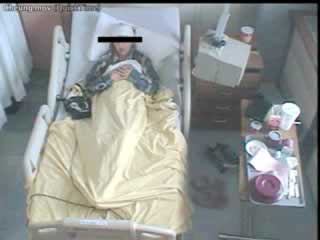
Upon admission to the epilepsy monitoring unit, continuous video-EEG monitoring confirmed the semiology of the events, but without clear EEG changes (see video sequence and figure 1). An MRI showed a right frontal transmantle cortical dysplasia extending from the right superior frontal gyrus down to the upper outer border of the right lateral ventricle (figure 2). An ictal SPECT showed hyperperfusion over the right frontal regions corresponding with the area of cortical dysplasia (figure 3). At this time it was felt that epileptogenesis was from the right frontal area and surgery was considered.
Intracranial electrodes were implanted and these showed a broad field (figure 4) corresponding to the obvious cortical, dysplastic lesion.
Resection of the right superior frontal tissue and white matter was performed and pathological analysis showed focal, cortical dysplasia with subcortical ballooned astrocytes.
She had been seizure-free for 12 months post-operatively and was driving again when she began to experience rare auras reminiscent of her previous seizures. These consisted of a brief, rising, abdominal sensation that was not associated with any change in level of consciousness, ability to speak or understand, and there were no associated involuntary movements. She continues to be on antiepileptic medications to control these auras.
Discussion
Malformations of cortical development are divided into three categories based upon the three major processes in cortical development: cell proliferation and apoptosis, neuronal migration, and cortical organization. Cortical dysplasia in particular is due to a defect in neuronal migration, with disruption of normal cortical lamination. Taylor type cortical dysplasias are focal epileptogenic abnormalities of the cerebral cortex containing dysplastic neurons within. These and various other types of dysplasias are characterized histologically by the presence of balloon cells (Taylor et al. 1971, Barkovich et al. 2001).In dysplasias where there are abnormalities of cell type from the time they are generated, radiological appearance reflects the inability of this cell type to migrate normally or organize correctly after arrival in the cortex. Thus, imaging should show abnormalities that extend from cortex to germinal zone near the ventricular margin (Barkovich et al. 2001) as would seem to be the case in our patient.
EEG investigation of patients with epilepsy due to Taylor-type cortical dysplasia has revealed distinctive ictal and interictal patterns characterized by disruption of background activity, high frequency fast spikes and polyspikes, interspersed with flattening and fast lower amplitude activity (Tassi et al. 2002). Focal, fast, rhythmic epileptiform discharges have also been found in patients with focal cortical dysplasia (Kuruvilla and Flink 2002. Gambardella et al. 1996). While present only in approximately half the patients studied, when present, such discharges would appear to be useful for identification of epileptogenic foci amenable to surgical removal. A review by Kutsy (1999) identifies several types of interictal patterns seen in patients with extratemporal epilepsy, including various focal, regional and generalized epileptiform discharges that may or may not localize to the site of seizure origin. Also noted is the distinct possibility that no epileptiform discharges may be seen at all, which was the case in our patient, likely due to her discrete area of activation. Scalp EEG findings are often poorly localizing (Raymond et al. 1995), necessitating further imaging to determine seizure focus.
SPECT brain imaging has proven helpful in localizing epileptogenic foci in patients with medically refractory epilepsy. The sensitivity of SPECT in localizing epileptic foci is highest for ictal, then post-ictal, then interictal recording, as noted in one meta-analysis (Devous et al. 1998). A similar pattern of sensitivity is noted for the relationship of SPECT to surgical outcome in the localization of seizure focus. In the case of our patient, the EEG was not helpful, and ictal SPECT was capable of revealing hyperperfusion over the right frontal regions corresponding to the area of cortical dysplasia seen on MRI.
Extensive imaging is often necessary to localize the lesion for epilepsy surgery as the completeness of resection correlates with degree of seizure-freedom (Alexandre et al. 2006). Even with extensive imaging however, focal cortical dysplasias are often missed on initial scan, and because they often occur concurrently with other pathology (Bautista, Foldvary-Schaefer et al. 2003), a consistent outcome is difficult to achieve. Malformations of cortical development can worsen the probability of complete resection as compared to hippocampal sclerosis or tumour, possibly because such malformations can extend microscopically beyond the resected region (Hirabayashi et al. 1993). Our patient was rendered completely seizure-free (Engel Class 1) after partial frontal lobe resection, which is consistent with Class I seizure relief obtained by 72% of patients with resection according to Kral et al. (2003), but somewhat unusual as the percentage of those seizure-free post-operatively tend to decrease progressively from childhood, through adolescence and adulthood (Edwards et al. 2000, Kral et al. 2003).
While gelastic seizures are commonly deemed a hallmark of hypothalamic hamartomas (Kerrigan et al. 2005), many studies have linked gelastic seizures to lesions of the temporal lobes, frontal lobes, pituitary tumours, astrocytomas of mamillary bodies, third ventricular papillomas, and various other locations (Loiseau et al. 1971, Ianetti et al. 1992, Arroyo et al. 1993, McConachie and King 1997). Our patient’s gelastic seizures were due to focal cortical dysplasia in the frontal lobe, not detected by EEG and identified only after MRI and SPECT localization. As frontal seizures, our patient’s episodes were also somewhat atypical in that they lacked prominent motor manifestations and presented instead with laughter and various sensations of restlessness or abdominal discomfort.
Finally, a possible explanation for the remaining auras in our patient is that perhaps not all the epileptogenic area was resected, as the changes during intracranial recordings were more regional than focal (Iannetti, 1992).
In conclusion, it is worth noting that gelastic seizures are not pathognomonic of hypothalamic hamartoma exclusively. Consideration should also be given other brain areas.


