Epileptic Disorders
MENUElectropositive seizures and non-convulsive status epilepticus in a critically ill patient with prior skull defect Volume 22, issue 2, April 2020
Epileptiform discharges captured on routine scalp EEG almost always carry a surface negative dipole. In rare instances, positive polarity discharges have been reported, usually caused by a skull defect (Fumisuke Matsuo, 1977; Franco and Kremmyda et al., 2018). In an intensive care unit (ICU) setting where critically ill patients undergo continuous EEG monitoring (cEEG), recordings are frequently marred by artifact. Given the rarity of surface positive discharges, adult electroencephalographers in the critical care setting may find it difficult separating artifacts from such cerebral potentials. Here, we present a novel case of a patient in non-convulsive status epilepticus (NCSE) where cEEG revealed frequent electropositive non-convulsive seizures.
Case study
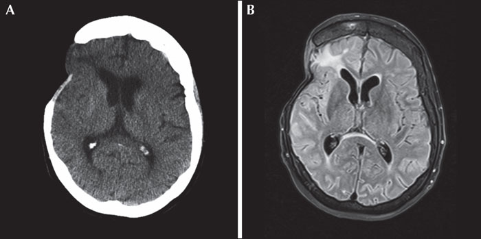
A 66-year-old woman was admitted to the neurology service following a generalized tonic-clonic seizure. Her history was significant for a right middle cerebral artery (MCA) aneurysm rupture with subsequent coiling. This was followed some years later by a second right MCA aneurysm coiling, complicated by thrombus and coil malposition which was salvaged with a stent. Subsequently, partial filling was noted in the first aneurysm and this was treated with a microsurgical clip. This was complicated by a bone flap infection requiring urgent removal during a recent admission. Medications on admission included Aspirin 81 mg daily and ramipril 10 mg twice daily. Initial investigations for seizure precipitants, including infections, toxins, metabolic changes and/or new structural lesions, were negative (figure 1A, B). She was started on phenytoin.
The patient was oriented but drowsy during the first days of admission. EEG performed on post-admission Day (PAD) 2 revealed continuous rhythmic spike, sharp wave and slow wave epileptiform activity arising from the right frontotemporal lobe, prompting treatment with levetiracetam for NCSE. EEG was repeated on PAD3 with no significant changes thus lacosamide was added, and on PAD4, the patient was transferred to the ICU for more aggressive anesthetic and anti-epileptic medication management of refractory focal NCSE.
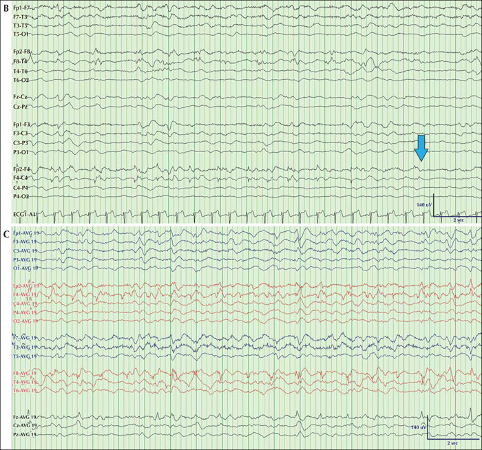
cEEG in the ICU revealed bilateral delta and theta slowing and lateralized periodic discharges over the right frontotemporal region with spatiotemporal evolution satisfying criteria for non-convulsive seizures; interestingly, the polarity was electropositive (figure 2).
Despite escalating anti-epileptic medications (including levetiracetam, lacosamide, topiramate, and clobazam) and alternating anesthetic regimens (including propofol and midazolam), the patient remained in NCSE for nine days in the ICU. She developed complications including ventilator-associated pneumonia, acute liver failure, and acute infarct in the right paramedian aspect of the pons. Discussion with the patient's family resulted in shifting focus to comfort care and she died the following day.
Discussion
We identified a previously unreported electrographic signature in a critically ill patient with predominantly surface positive non-convulsive seizures satisfying criteria for NCSE. The signature itself was unassuming, characterized mainly by low-voltage arrhythmic fast activity with intermixed electropositive spikes emerging from a disorganized and attenuated background rhythm, the latter owing to the sedative medication regimen the patient was treated with in the ICU setting. Critical care EEGs are frequently captured in an environment propitious to recording artifacts and the observed pattern could have been mistaken for electrode artifact over F4/F8. However, several clues revealed the malignant nature of the signature itself: the interictal surface negative sharp waves over F4, the focal polymorphic theta activity over F4/F8, and only the rare but informative appearance at ictal onset of surface negative lateralized periodic discharges over F4 (figure 2A). Additionally, the electrographic signature revealed delta activity over F4, identifying a clear end to the pattern (figure 2B).
Epileptiform discharges are typically surface negative owing to the perpendicular orientation of dipoles relative to the cortical surface and to scalp electrodes (Tharp, 1971; Goldensohn, 1975). While electropositive spikes and sharp waves are considered rare in adult populations, they are common in neonatal and pediatric populations, owing in part to the incomplete migration of cortical neurons as well as immature myelination. They are predominantly seen in neonatal critically ill patients, in benign epilepsy with centrotemporal spikes (Luders and Lesser, 1987; Kellaway, 2000) or in patients with structural abnormalities such as interventricular hemorrhage or periventricular leukencephalomalacia (Gregory and Wong, 1992). Rare instances in adults have been reported in patients with skull defects and breach rhythms (Fumisuke Matsuo, 1977; Franco and Kremmyda et al., 2018). Lee et al. identified their presence in patients with breach rhythms undergoing magnetoencephalography (MEG) to better characterize epileptiform activity inside the area of breach (Lee et al., 2010). Three patients had surface positive sharp waves on the scalp EEGs, two of which also showed interictal epileptiform discharges on MEG recordings, thus suggesting their epileptiform potential.
The notion that surface positive sharp waves are associated with seizures and epilepsy was reinforced by Franco et al. who retrospectively studied 24,178 EEG reports from 18,060 separate patients and identified eight recordings (0.033%) with electropositive sharp waves, most commonly seen in patients with prior brain surgery (Franco and Kremmyda et al., 2018).
Our patient had a prior history of right frontal craniotomy and encephalomalacia which altered her cortical anatomy and likely explains the unique surface positive dipole observed while recording her non-convulsive seizures. To our knowledge, this is the first report of an electropositive non-convulsive seizure in the adult critically ill/continuous EEG monitoring literature.
Surface positive epileptiform abnormalities are rare in adult populations and predominantly observed in patients with prior brain surgeries and skull defects; they are known to be associated with seizures and epilepsy. Here, we report on a critically ill patient with a prior right frontal craniotomy whose cEEG captured surface positive non-convulsive seizures and NCSE that could easily have been attributed to electrode artifact given their morphology and polarity. In the ICU setting, where cEEG recordings are frequently confounded by artifacts, the electroencephalographer should interpret surface positive discharges with care and refrain from underestimating their epileptogenic nature.
Acknowledgements and disclosures
We would like to thank Dr. Denise Cook for her contributions in the editing of this manuscript.
Dr. Fantaneanu has received speaking honoraria from Sunovion, Eisai and UCB and a travel grant from Eisai. None are relevant to this case.


