Epileptic Disorders
MENUEarly Parkinsonism in a Senegalese girl with Lafora disease Volume 22, issue 2, April 2020

Figure 1
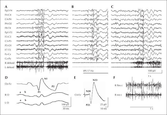
(A-C) EEG samples showing diffuse polyspike discharge followed by diffuse slow waves, associated with ictal myoclonia-atonia (A), photoparoxysmal response to intermittent photic stimulation (IPS, at 17 Hz) (B), and multifocal and diffuse spikes (C). (D) Flash-VEPs (top trace), and ERG responses (lower traces). Note increased amplitude of VEP responses and attenuated ERG a and b waves. (E) SEPs. Note giant P25-N33 response. (F) EMG sample from upper limb muscles showing low-amplitude and short myoclonic bursts.

