Epileptic Disorders
MENUBilateral occipital dysplasia, seizure identification, and ablation: a novel surgical technique Volume 16, issue 2, June 2014

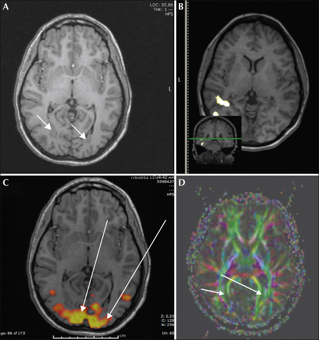
Figure 1
(A) Bilateral cortical dysplasia extending into both occipital lobes (greater in the right side than left) from both peri-ventricular regions (arrows). (B) Subtraction SPECT demonstrated hyper-perfusion involving the right posterior temporal, occipital, and inferior parietal regions. (C) Functional MRI with bilateral bold signals hugging the regions of cortical dysplasia (arrows). (D) DTI/tractography revealing tracts which separate and surround the dysplasic tissue in both occipital lobes (arrows).

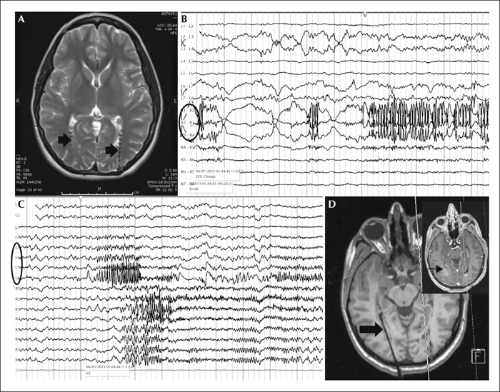
Figure 2
(A) Bilateral depth electrodes lateral to, and partially traversing regions of, dysplasia. (B). Seizure onset and evolution involving the right (R) depth electrode (circle). (C) Seizure onset involving the left (L) depth electrode (circle), then rapidly spreading to the right. (D) Ablation catheter (thick arrow) and area ablated (thin arrow).

