European Journal of Dermatology
MENULeg ulcers associated with cutaneous vascular degeneration in a patient receiving pazopanib chemotherapy Volume 27, issue 5, September-October 2017

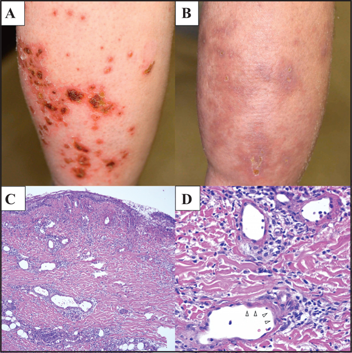
Figure 1
Clinical and histopathological features of the ulcers. A) Multiple skin ulcers with crusts on the flexor side of the lower legs. B) The ulcers improved two weeks after pazopanib discontinuation. C) Histopathological features of a skin biopsy specimen from an ulcer on the right leg. An open ulcer and dilated blood vessels with inflammatory cell infiltration; degeneration of the vessel wall is observed. D) Oedematous change and pyknosis of endothelial cells on the vessel walls (white arrowhead), but no apparent infiltration of inflammatory cells. Hematoxylin-eosin staining; original magnification: ×100 (C) and ×200 (D).

