Epileptic Disorders
MENUThe neural networks underlying the illusion of time dilation: case study and literature survey Volume 24, numéro 3, June 2022
Illustrations
It has been over a century since Einstein showed time as a mere measurable dimension of spacetime susceptible to speed. The twin travel paradox is no less fascinating, that no specific organ exits for time sense akin to the ears and eyes for sound and vision. Time is rooted in the survival of many species. The essential part of time perception relates to the natural circadian cycle and the seasonal transitions related to the earth’s yearly rotations around the sun. The ever-accelerating pace since the industrial revolution is an excellent example. Humans’ unique linguistics, time perception, and abstract skills distinguish our cultural evolution from other species. Surely, time has been important to every civilization. What has emerged is our perception of time does not equate with an objective reality external to ourselves. Evident, for instance, in persons undergoing anesthesia or emerging from a coma. The sense of time passing is instead, generated within our brains through the perception of the environment’s temporal features. The brain mechanisms underlying this temporal processing, however, are not delineated.
To that end, we report on a profound illusionary alteration of time and duration perceptions in an epilepsy patient during direct electrical cortical stimulation for functional mapping. We analyze the cortical and subcortical networks relating to that experience. To our knowledge, this account is the first original of a human’s experience of time dilation during direct cortical stimulation.
Material and methods
Please refer to supplementary material for details. Briefly, the patient was undergoing invasive EEG evaluation to localize the epileptic focus at Yale Comprehensive Epilepsy Center. Electrode implantations and 50-Hz electrical stimulation via 3-5-second trains for mapping function were performed per standard of care. Granger causality and phase-locking value for each electrode pair were calculated using high-frequency oscillations greater than 80 Hz. Functional MRI connectivity analysis was performed on BOLD signal via arithmetic correlation. The patient approved participation in the study.
Case presentation
A 56-year-old, right-handed man with a nine-year history of refractory focal aware seizures and rare focal-to-bilateral tonic-clonic seizures presented for surgical evaluation. Typical attacks consisted of arousal from sleep, a wave of rushing sensations over all of the body, respiratory difficulty, loud vocalization, and drooling with left facial motor twitching and rarely secondary generalization. The typical duration was 30 seconds to 2 minutes. The seizures were poorly controlled with anticonvulsants. Risk factors included a history of meningitis within childhood, an older brother with seizures, and head trauma secondary to contact sports with brief loss of consciousness. There was a remote history of cocaine and lysergic acid diethylamide (LSD) usage in teenage years. The neurological examination was unremarkable. The neuropsychological testing showed high average to very superior performance across multiple domains with a full-scale IQ of 125. Long-term non-invasive video-electroencephalogram revealed isolated and periodic epileptiform discharges in the right centrotemporal regions. Ictal video-EEG recording during left lower facial twitching and left head jerking also revealed frequent epileptiform discharges in the right centro-temporal regions. Brain magnetic resonance imaging (MRI) was normal. The functional MRI (fMRI) showed left hemispheric dominance for language. The patient subsequently underwent intracranial electrode evaluation for localization of the epileptogenic region and functional mapping and localization for face and hand regions over the right hemisphere.
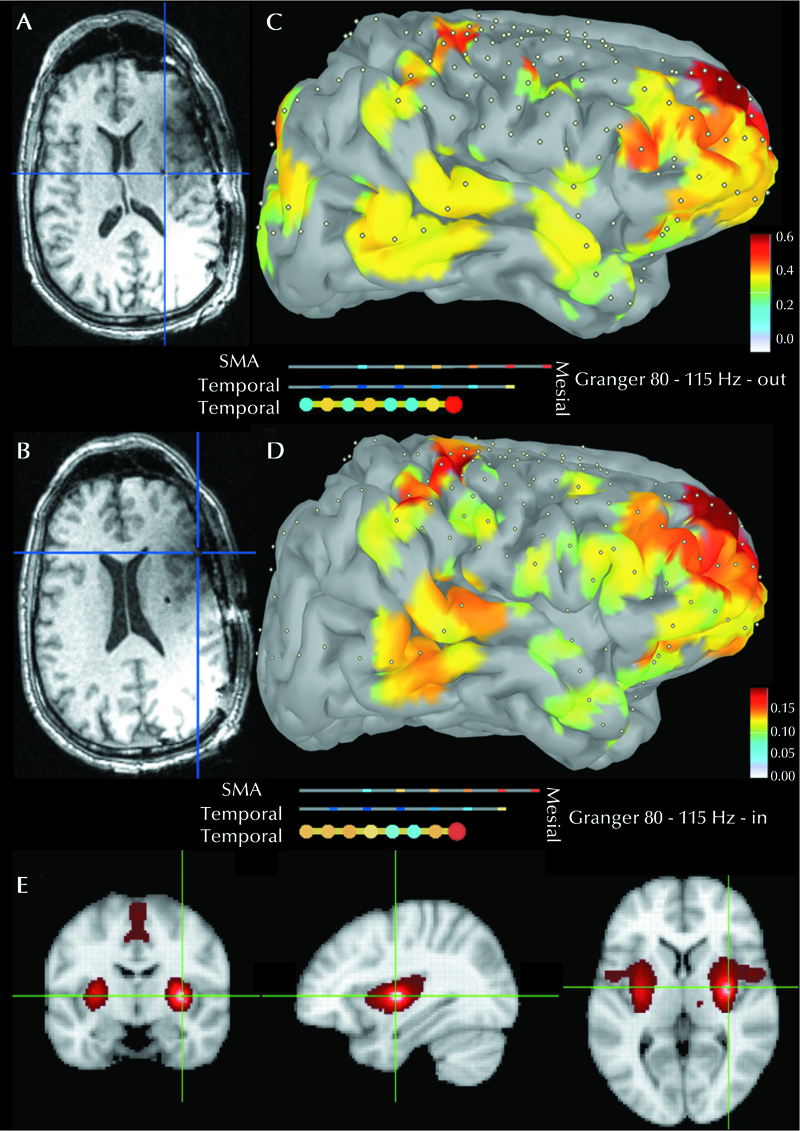
The implantation spanned the right hemisphere and the orbito-frontal region over the left. Different sensorimotor functions were recorded in 46 stimulation sessions (median: 4 mA; interquartile range: 3-6 mA) or no functions were recorded (mode: 12 mA). The stimulation of contacts implanted in the right claustrum (figure 1A, supplementary figure 1), with a contact at the level of mid-dorsal right insula (4 mA), consistently correlated with the psychic experience described by the patient as “felt like an eternity”. The patient indicated that he had an insight of what might have been the actual duration inferred from the stimulation of other sites, however, he emphasized that it was perceived as substantially longer; “hours” per estimate, nonetheless. Longer trains of 5 vs. 3 seconds correlated with longer interpretation of that interval without the ability to quantify further. The patient was asked to recite the ascending series of digits during stimulation (5-7, timed subjectively at 1 digit per second) and subsequently again confirmed the same symptoms. The patient was able to move, name and speak during stimulation. He passed standard language tasks. He denied pain. He attributed his apparent discomfort to the distraction resulting from the insight into the time perception illusion. The patient recalled specific phrases provided during stimulation and denied correlation with seizure symptomatology or auras, though later divulged that the symptoms overlapped with that experienced while “tripping” as a teenager with LSD and mushrooms. Less intense symptomatology was observed with higher current stimulation over the right pars opercularis (figure 1B) in the inferior frontal gyrus (9-11 mA). The symptoms were not reproducible with three sham stimulations at each site. No afterdischarges were noted during or after the stimulations. The finding was reproducible on the second day of the stimulation mapping session. The sites exhibited bidirectional connection, with a stronger outflow component with the immediate vicinity, right superior frontal gyrus including mesial aspects, mesial temporal and to less extent the right intra-parietal region, prefrontal area at large, and the temporo-occipital junction.
Phase-locking values showed consistent connections with the right superior frontal gyrus and parsopercularis. Resting-state functional-MRI revealed co-activation in the bilateral basal ganglia, contralateral cerebellum, and bilateral insula and pre-motor regions (figure 1C-E). The patient underwent a resection of the right face motor area, where the seizure onset was recorded, and seizures were induced by stimulation, sparing the regions producing the symptoms. The patient remained free of motor seizures following the resection at the 27-month follow-up visit.
Discussion
The stimulation of the right claustrum/dorsal-mid insula and the non-dominant right inferior frontal regions, homologous to Broca’s area, corresponded with a perception of time dilation. Unlike other senses with dedicated organs, the matter is complicated with time sense. To our knowledge, this is the first report of subjective time dilation produced by electrical cortical stimulation. The scarcity of such reports is likely not fortuitous. It is most consistent with a right hemispheric “non-dominant” lateralization, not typically mapped for clinical purposes. It is compatible with records of right hemispheric preference for time perception based on correlations with structural lesions in a human and electrophysiological investigations [1]. Time judgment is essential for many living species in which short-timescale, temporal information plays a crucial role including, e.g. estimation of how long to look away in peace, sophisticated pauses in speech and what these may convey, to the rigor of execution of motor movements and decision making. We propose the stimulated areas may correspond to a pacemaker of the internal clock in the order of seconds/minutes according to the psychological models of scalar time theory. The pacemaker accumulator model and the extension of that, the attentional gate model, suggest that an internal clock generates regular pulses, and an accumulator keeps track of these pulses. There is a view of multiple pacemakers depending on the timeframe of duration; from those relating to circadian rhythms (e.g., optic pathways/ hypothalamus) to the basic motor decision in the order of milliseconds (e.g., basal ganglia, SMA and cerebellum), to a higher degree of cognitive perception and complex decisions of longer durations where the insula and pre-frontal regions correspond with more advanced human behaviors and the formation of episodic and working memories. Those different time scales and hierarchies correlate with reports of differential effects of drugs on time judgment such as those seen with benzodiazepines compared to haloperidol. Time perception in that prospective sense differs from that retrospective one likely judged by memory, recall, and relationship to other events [2]. The electrophysiological connectivity profile with the superior frontal gyrus correlates with the complexity of the executive relationship, which is most developed in the human brain compared to other species. The relationship with the mesial frontal and the supplementary motor areas highlights the expected involvement of time processing in motor decision-making. This was further evident with fMRI connectivity also showing cerebellar and basal ganglia involvement. The correlation with the intra-parietal and temporo-occipital regions is consistent with time interpretation in the sensory stimuli context. Thus, there is little to no time appreciation in coma, under sedation, or during sleep, suggesting a strong correlation with consciousness per se. The relationship to the mesial temporal structures is compatible with the recent discovery of time-cells correlating with the sequence of observed stimuli and the formation of the episodic memory, recently confirmed in humans as well [3]. The time-cells oscillate when place/grid cells are idle, interestingly, at paces modulated by running velocity in an empirical model to allow relative phase to code space. Longer consolidations involve the insula according to functional imaging surveys [4]. The insula has a higher proportion of Von Economo neurons. These cells are believed to have the unique function of making the association between emotions and actions. Just like manual dexterity is dependent on close connections between contiguous somatosensory and motor areas, the Von Economo neurons are believed to enable fast integration of emotions and behaviors. Degeneration of these cells in frontotemporal dementia is believed to result in loss of emotional awareness and self-conscious behaviors that are typical of that condition. These neurons are not found in macaque monkeys, and they are fewer in chimpanzees and gorillas than inhumans. Interestingly, they are more numerous in adult humans than in infants, which suggests that their proliferation parallels the accumulation of affect-encoded knowledge across the lifespan. It ispossible that any divergence fromprior reports of bi-hemispheric involvement in functional imaging stems from the unresolved issue of scaling of statistical thresholds. The mapped networks are compatible with disorders showing the most pronounced alteration in time awareness such as in ADHD, impulsiveness, Parkinson’s disease, and schizophrenia [5], or even in fluctuating of states of mind, such as in boredom or the subjective sense of “fun”. The role of the dopaminergic system is evident with alterations encountered in drug use, Parkinson’s and with aging. There is no evidence, however, that dopaminergic replacement in Parkinson’s disease reverses alterations of time perception.
Transcranial stimulation studies did not produce consistent findings in mapping time networks [6], owing to differences in stimulation, and no reliable testing paradigms, unlike the findings with electrical stimulation, not only correlating with spatial precision but with functional precision akin to that required for the surgical resection planning. One could view that loss of consciousness is the ultimate outcome of induced low-pass filtering of time perception, as recently reported by stimulation of a congruent region on the contralateral side in humans [7]. However, those, along with other reports of ecstatic phenomena produced by electrical stimulation of the dorsal insula [8, 9], should be interpreted with caution considering the relation to the epileptic network, lateralization, stimulation parameters, and anatomy specifics. For example, the site of stimulation in our case was not involved in epileptiform discharges and seizure spread. There is a suggestion that time is perceived via causality. More generally, time is an aspect of the relationship between events. Since the claustrum has been hypothesized to bind multimodal sensory input into what we perceive as a single experience, disrupting its function is compatible with altered time perception.
In conclusion, the findings of this case are agreeable with the hierarchal view of time perception in humans. At the highest level, it depends on integrating multiple neural systems emerging from various sensory and executive networks, in which the right mid-claustrum/ insula and the inferior frontal lobe regions may play the role of a pacemaker interacting with the accumulators spanning large neocortical and mesial temporal regions. To our knowledge, this phenomenon has not been reported in humans before in these settings. In the physical sense, however, time will continue to march regardless of one’s own perception.
Supplementary material.
Supplementary data accompanying the manuscript are available at www.epilepticdisorders.com.
- (1)Is there hemispheric preference in time perception?
- (2)Since there are no dedicated organs for time perception in the body and brain, how would you interpret the findings in the report?
- (3)Based on that, is time perception relative in the brain as well?
Disclosures.
The authors have no conflicts of interest to declare that are relevant to the content of this article.


