Epileptic Disorders
MENUStimulation-induced ictal vocalisation of left frontal lobe origin Volume 20, numéro 5, October 2018


Pure ictal non-speech vocalisations have been observed in selected patients with frontal and temporal lobe epilepsies (Janszky et al., 2000; Rego et al., 2006; Bonelli et al., 2007; Horvath et al., 2009). Previously, we reported pure ictal vocalisation in a patient with left frontal lobe epilepsy (FLE) (Rego et al., 2006). In this report, we present another patient with FLE in whom we consistently elicited monotonous vocalisation by electrical stimulation of the white matter of the left superior frontal gyrus.
Case study
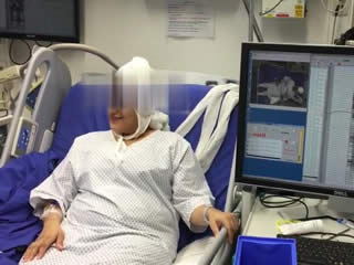
A 31-year-old, right-handed female with medically refractory focal epilepsy of the left anterior insula since the age of 12 was admitted for invasive EEG monitoring with depth electrodes (Patient 1). Despite antiepileptic drug treatment, she continued to have motor seizures mainly during sleep with an average frequency of 2-3 per week. Her past medical history was unremarkable. Family history revealed a brother with pharmacoresistant focal epilepsy who was seizure-free after resective epilepsy surgery. Her neurological examination was normal. Cranial MRI did not reveal any lesions. She underwent non-invasive continuous EEG monitoring to evaluate the option of epilepsy surgery which showed either frontal non-lateralized or left frontal seizure onset. Interictal non-invasive EEG revealed no interictal epileptiform discharges. Interictal 18-fluorodeoxyglucose-positron emission tomography (FDG-PET) revealed hypometabolic areas in the left insula as well as in bilateral parietal, temporal, and central regions. Ictal single-photon emission computerized tomography (SPECT) (neurolite 99mTc) showed hyperperfusion in the left inferior temporal and left orbito frontal regions. Diffusion tensor imaging (DTI) demonstrated U-fibre density reduction in the left frontal area (figure 1A). Invasive EEG evaluation with stereotactically implanted depth electrodes was performed to localise the seizure onset zone and to outline the eloquent cortex (figure 1B). The exact localisation of intracranial electrodes was defined by fusion of 3-dimensional reconstruction post-implantation computed tomography (CT) scans and preoperative MR images. The invasive evaluation localised the seizure onset in 31 seizures to the left anterior insula (H 2-6) which propagated into the left frontal lobe (D 5-8, F 5-7) within a few seconds. Maximum interictal epileptiform discharges were located mainly in the left anterior insula (H 1-4) and, to a lesser extent, in the left superior (A, C, D) and middle frontal gyrus (F) (figure 1C). Extraoperative electrical stimulation of the left superior frontal gyrus (50-Hz electric current in square waves with a 5-mA intensity and a duration of 5 seconds, delivered at left medial superior frontal gyrus electrodes C6-C8, with monopolar stimulation), without after-discharge, consistently elicited a monotonous vowel lasting for the duration of stimulation (figure 2A, video sequence 1). Subtle retraction of the head followed the vocalisation. The results of electrical stimulation at all electrodes are illustrated in figure 1C. Neurosurgical resection of the left anterior insula and the superior and medial frontal gyrus was planned.
We were puzzled by the similarity of the vocalisation elicited by electrical stimulation between this patient and a former patient who had left FLE with pure ictal monotonous vocalisations reported several years ago (Rego et al., 2006) (Patient 2) (video sequence 2). The stimulation-induced vocalisation was not part of the habitual seizures of Patient 1. Quantitative analysis of audio signals was performed in both patients. Quantitative analysis leads to a description of the frequency of peaks of a sound, which are called formants. Audio signals were recorded with Shure MX202 microphones (Shure, Niles, IL, USA), using the uncompressed pulse code modulation format and a bitrate of 64 kbit/s, and saved as waveform files. Quantification of the pitch (f0), i.e. the fundamental vocalisation frequency in Hertz, was obtained by using the pitch listing tool of the audio-analysis software, Praat (Version 6.0.05, freeware, www.fon.hum.uva.nl/praat/; ©1992-2015 by Paul Boersma and David Weenink, University of Amsterdam, Netherlands [Boersma and Weenink, 2015]). Regarding the audio analysis of Patient 1, the mean fundamental frequency (f0) of the vowelwas higher and showed considerably less frequency variations (expressed as standard deviation of frequency) for the stimulation-elicited voice compared to her voice without stimulation (mean pitch frequency: 213.5 Hz±5.3 vs 207 Hz±27.3, respectively) (figure 2A). Regarding the audio analysis of Patient 2, the mean fundamental frequency (f0) of the vowel, which was produced ictally, was higher and the frequency variation was lower compared to the interictal state (mean pitch frequency: 184.6 Hz±19.2 vs 172.1 Hz±62.1, respectively) correspondingly (figure 2B).
Discussion
In this report, we show that pure ictal vocalisation in a patient with FLE, which we published 12 years ago (Rego et al., 2006), sounded very similar to electrically evoked non-speech monotonous vocalisation in another patient. Pure ictal vocalisations, defined as an audible non-speech sound, not accompanied by apnoea or generalized tonic-clonic or clonic seizures (including groaning, grumbling, shouting, and weeping sounds), have been reported in patients with FLE with conflicting results on lateralization (Janszky et al., 2000; Bonelli et al., 2007). However, ictal vocalisations corresponding to any sounds without speech quality, as part of any seizure type, have also been observed in dominant temporal lobe epilepsy (Gabr et al., 1989; Horvath et al., 2009). Spread of the ictal activity from the temporal to the frontal lobe has been speculated in these patients to be responsible for ictal vocalisation (Gabr et al., 1989; Horvath et al., 2009).
The electrodes from which stimulation elicited the similar sound to the ictal vocalisation of our former patient were located in the superior frontal gyrus (Rego et al., 2006). This observation is, therefore, suggestive of a symptomatogenic zone of vocalisation in the left superior frontal gyrus. Our results are in concordance with previous reports that non-speech vocalisation is evoked by electrical stimulation of predominantly the left superior frontal gyrus (supplementary motor area [SMA]); a sound described that was similar to those in our patients (Penfield and Welch, 1951; Fried et al., 1991; Lim et al., 1994). Former studies have identified representation of the human laryngeal motor cortex in the primary motor cortex (area 4) (Penfield and Boldrey, 1937). However, functional imaging studies showed a widely distributed network including the medial SMA, anterior cingulate cortex, and motor and premotor regions in the left hemisphere producing vowels and syllables (Loucks et al., 2007; Simonyan et al., 2009).
Our approach is new in the sense that we analysed the audio signal quantitatively, showing similarity between the vocalisations. Quantitative analysis of the evoked vocalisation (by electrical stimulation of the left superior frontal gyrus) of the first patient demonstrated a higher mean fundamental frequency (f0) of the vowel and a decreased alteration of the variation of the pitch compared to spontaneous voice, which is similar to the ictal vocalisations of the second patient with left FLE (Rego et al., 2006). In a recent study, more prominent increases in ictal f0 were identified during seizures arising from the frontal lobe compared to those from the temporal lobe, suggesting frontal lobe involvement in laryngeal control and speech production (Speck et al., 2018). In addition, ictal alteration of vocal prosody, as shown by change in the frequency of formant f0 and a reduction in frequency variation during speech, was found mostly in seizures arising from the non-dominant temporal lobe (Peters et al., 2011). Quantitative voice analysis contributes to objective evaluation of ictal and postictal vocalisations in epilepsy patients (Peters et al., 2011).
Since the symptomatogenic zone of ictal vocalisation is overlapping with some negative motor areas with subsequent arrest of motion arising from prolonged electrical stimulation of the left mesial frontal area (SMA) (Ikeda et al., 2009), we also elicited negative motor responses in the regions adjacent to those where electrical stimulation caused pure vocalisation in the left superior frontal gyrus (C1). The role of other regions adjacent to stimulated cortex cannot be excluded due to limitation of sampling with depth electrodes in our patient.
Conclusion
We conclude that continuous monotonous prolongation of a vowel as the sole manifestation of an epileptic seizure can originate from the left superior frontal gyrus. This phenomenon should be further evaluated with quantitative audio analyses as this may be potentially useful for presurgical evaluation of epilepsy patients.
Supplementary data
Summary didactic slides are available on the www.epilepticdisorders.com website.
Acknowledgements and disclosures
Dr Baysal-Kirac was supported by the International Federation of Clinical Neurophysiology.
None of the authors have any conflict of interest to declare.