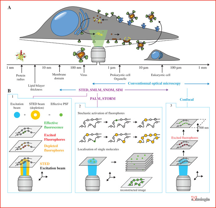
Size scales visible by different conventional or super-resolution optical microscopy techniques.
A. Schematic representation of an infected eukaryotic cell, and the different sizes of biological objects observable by the corresponding microscopy techniques. B. Operating principle of STED, PALM/STORM, and confocal microscopes. 1. Stimulated Emission Depletion Microscopy (STED) consists in bringing excited fluorophores back to their fundamental (non-fluorescent) state via a non-destructive process: the excitation laser puts the targeted fluorophores in their excited state and then, a red-shifted laser (called STED laser) is added and returns the already excited fluorophores to the non-fluorescent state. By modulating the focal intensity distribution of the STED laser so that it has at least one intensity minimum (e.g. a donut-shaped intensity distribution), fluorescence is depleted everywhere except at the local minimum (effective PSF up to 20-30 nm in diameter). 2. SMLM microscopies allow the detection of single molecules labeled with photo-convertible fluorophores. These techniques are based on image reconstruction from a set of 100-10,000 individual acquisitions where only small subsets of isolated fluorophores are stochastically activated. The spatial coordinates of the diffraction spots centers produced by these fluorophores are accurately determined in all acquisitions, which are then used to reconstruct the final image. Depending on the technique, the stochastic on/off switching of fluorescent markers is achieved by different means: PALM microscopy controls the fluorescence of photo-convertible molecules (e.g. PA-GFP, PA-mCherry, mEOS, mMAPLE, etc.) by short stimulations. In response, the molecules emit light one after the other (“blink”), and it is then possible to separate the photons coming from each emitting molecule. STORM microscopy is a technique based on the same principle, but instead uses organic markers such as paired cyanines. Both techniques are applicable to living biological systems. 3. Confocal microscopy, also based on the excitation of fluorescent markers, employs a device capable of blocking the out-of-focus fluorescence and thus capturing only the focal plane fluorescence. Images of minimal depth of field (about 400 nm) called “optical sections” are therefore obtained. NSOM: Near Field Scanning Optical Microscopy; SIM: Structured Illumination Microscopy; PSF: Point Spread Function.
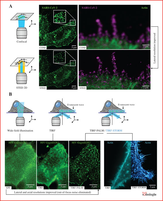
Comparison of different microscopy techniques applied to the observation of human viruses: From classical optical microscopy to super-resolution microscopy.
A. Comparison of images obtained by confocal microscopy versus stimulated emission-depletion microscopy STED. Left panels: VeroE6 cell infected with SARS-CoV-2 coronavirus (green). Right panels: Zoom on the membrane of a VeroE6 cell infected by the SARS-CoV-2 coronavirus (in magenta) and labelled for cellular actin (in green). Virus size = about 100 nm in diameter. STED microscopy enables to obtain a better resolved image and to distinguish single viral particles. It allows to overcome the diffraction limit imposed by the wave nature of light and to reach a lateral resolution lower than 50 nm (where confocal microscopy does not allow to go below 200-250 nm). B. Left panels: HEK 293T cells transfected with HIV-1 viral protein Gag fused to the photoconvertible mEOS2 protein (green to red). Comparison of images obtained by wide-field fluorescence microscopy, TIRF microscopy, and photo-activated localization microscopy PALM (coupled to TIRF microscopy). As opposed to epifluorescence microscopy which illuminates the entire sample (including outside the focal plane), total internal reflection fluorescence (TIRF) microscopy permits selective excitation of the sample. In fact, the orientation of the incident beam is adapted to produce an evanescent wave (in blue) illuminating only a thin optical section of the sample surface (less than 200 nm). This improves the axial resolution, eliminates the out-of-focus fluorescence noise, and thus provides a better signal to noise ratio. The coupling of TIRF microscopy with PALM super-resolution microscopy (allowing to randomly activate some fluorophores and then to “turn them off”) finally enables to acquire an image below the diffraction limit, reconstructed after having determined with precision the position of all individual molecules. Right panels: HeLa cells labeled for cellular actin (in blue). Comparison of images obtained either by TIRF (left) or by a TIRF - STORM coupling. Here, STORM microscopy, which is based on the same principle as PALM (the difference residing essentially in the type of fluorophores used), can reach a resolution of 16 nm (against 250 nm for TIRF).
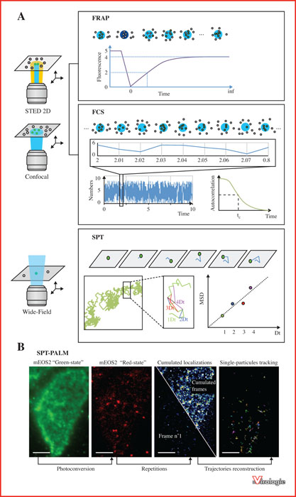
Single molecule and particle dynamics microscopies using FRAP, FCS and SPT.
A. FRAP: Fluorescence Recovery After Photobleaching. A technique that locally quenches photo-destructible fluorophores in an irreversible manner using a very intense and brief light beam. The return of fluorescence in the quenched area, due to the mobility of the molecules, is then analyzed using kinetic models. This allows to obtain quantitative and qualitative information on the nature of the molecules’ movement in the photo-destroyed zone. FCS: Fluorescence correlation spectroscopy. This technique is based on the analysis of temporal fluctuations of fluorescence in a given area of the sample. In this approach, we record the fluorescence emitted in the confocal or STED volume containing only a small number of particles. The fluctuation of the fluorescence intensity reveals the variation of the number of molecules present in the observed volume (concentration). The temporal autocorrelation of these fluctuations provides an average residence time of the molecules in the observed area, which gives information on the size and nature of the molecules’ movement. Hence, the analysis enables to obtain the average number of particles (concentration) but also the characteristic time of the movement. SPT: Single-particle tracking. This method is based on the analysis of the movement of individual particles in a medium. The time series of spatial coordinates, in 2D (x, y) or 3D (x, y, z), is called trajectory. The trajectory is typically analyzed using statistical methods to extract information about the underlying dynamics of single particles (by plotting the MSD, Mean Square Displacement, as a function of time). In particular, these dynamics can reveal direct information about the medium in which the particle is moving. In the case of random motion, trajectory analysis can be used to measure the diffusion coefficient. B. Example of the application of SPT coupled with PALM microscopy. HEK 293T cells transfected with HIV-1 Gag protein fused to the fluorescent tag mEOS2. From left to right: native “green form” of mEOS2; “red form” of mEOS2 after photo-conversion (stochastic activation of these fluorophores allows to define and precisely follow their localization); cumulative localizations of stochastically activated single mEOS2 molecules; reconstruction of single molecule trajectories over 100 images (time between each image = 22 ms). Scale bars = 5 μm.
La révolution très récente de la microscopie optique, associée à l’optimisation de la résolution spatiale et de la vitesse d’observation, a ouvert l’accès à la visualisation d’objets nanométriques. Ainsi, l’utilisation d’une nouvelle génération de microscopes de super-résolution combinée à l’amélioration des marqueurs fluorescents pour les protéines ou les lipides, permet désormais d’étudier le cycle réplicatif des virus à l’échelle de la molécule unique dans des modèles cellulaires fixés, mais aussi vivants, avec une précision nanométrique. Après une brève description chronologique et technique de ces nouvelles approches, nous détaillerons dans cette revue quelques exemples de microscopies de super-résolution ayant permis de revisiter notre compréhension du cycle de réplication de certains virus affectant l’humain, dans l’étude des relations hôte-pathogènes à l’échelle du virus et de la molécule unique.