Epileptic Disorders
MENUSeizure semiology: ILAE glossary of terms and their significance Volume 24, numéro 3, June 2022
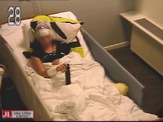
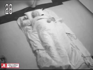
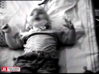
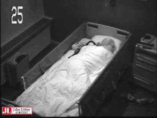
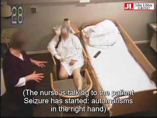
Seizure semiology comprises objective signs and subjective symptoms reported by patients during epileptic seizures. Accurate delineation of seizure semiology is essential for properly diagnosing and classifying seizures and epilepsy syndromes. Although valuable information can be obtained from the patients, and from people who have witnessed seizures [1], more detailed feature-extraction is possible by reviewing video recordings of the typical events [2]. Recent technological advances with high-resolution video cameras allow recording of subtle details, such as goosebumps, flushing and pallor. Combining EEG that is synchronized with the video allows detailed analysis and sequential electroclinical correlation from seizure onset to seizure termination.
Information obtained from patients concerning subjective seizure symptoms (not visible on the video recordings) are essential for correct characterization of the seizures, and for correct localization and lateralization, as described in detail in a previous seminar in epileptology [1]. The term aura denotes subjective symptoms at seizure onset that are perceived by the patient. It has been described in focal seizures (i.e., focal aware seizures and at the onset of focal impaired awareness seizures, before impairment of consciousness). Aura has also been reported in generalized seizures. Epileptic aura must be differentiated from aura occurring in non-epileptic paroxysmal events (migraine, syncope, dissociative seizures).
Our goal is to provide a list of terms that are used to describe seizure semiology, and to update the glossary initially developed in the ILAE Commission report published 20 years ago [3]. Semiology features were subdivided to reflect similar signs and their corresponding location. We acknowledge that other categories are possible. However, our focus was not to classify the seizure semiology, but rather, to describe the individual characteristics according to a commonly accepted terminology, that may be used individually or in combination to aid in defining the ictogenic network. For further information regarding definitions and classification of seizures and epilepsy, the reader is referred to the ILAE position statements [4-6].
We also included phenomena that occur immediately after seizure termination to distinguish postictal semiology and emphasize their clinical relevance. We used modifiers in accordance with what was previously proposed [7]. For some features, the terminology used to describe semiology applies to the body part that is involved (for example “epigastric aura”), and in other cases somatotopic descriptors have been added (for example “left hand gestural automatisms”). Other descriptors of seizures including duration, timing, and provoking or facilitating factors are also listed and defined.
This glossary of terms was designed to address learning objectives outlined by the ILAE curriculum and encompass an education review. Section 1.3.3 of the ILAE curriculum includes “Extract semiology information from video recordings” and section 1.3.4, “Interpret semiological signs and symptoms allowing hypotheses on the localization of focal seizures”[8]. This report has been developed by a working group of the ILAE Commission on Diagnostic Methods involving the EEG Task Force. For each semiology feature we used the following search strategy in PubMed: (((name of the semiology feature [Title/Abstract]) OR (synonym widely used [Title/Abstract])) AND ((localization [Title/Abstract]) OR (lateralization [Title/Abstract]))) AND (seizure [Title/Abstract]). Date for last search was 1/11/2021. In addition, we included papers from citations found in reference lists and review articles. Table 1 summarizes the most plausible localization and lateralization accepted for the most important semiological features. Table 2 lists video examples.
1. Electrographic seizures
Electrographic seizures are subclinical and do not have any observable signs or reported symptoms despite an unequivocal ictal pattern on EEG: 1) sustained spike-wave discharges with a frequency ≥ 2.5 Hz; 2) rhythmic – evolving ictal activity (with a dynamic evolution in time and space). Electrographic seizures (10 seconds and longer) and electrographic status epilepticus (10 minutes and longer) are often recorded in critically ill patients [9-12], and also in genetic or developmental / epileptic encephalopathies (such as patients with ring chromosome 20, Angelman syndrome and related genetic syndromes), in neonatal epilepsies [13] and in acute symptomatic neonatal seizures.
2. Consciousness
Impaired consciousness is of major clinical importance in epilepsy, affecting the ability of patients to safely drive, effectively work, and maintain the ability to function in society [14-17]. However, because the term “consciousness” is broad, the 2017 ILAE classification adopted the narrower term “awareness” in the classification, as a “simple surrogate marker for consciousness” [5]. Another related feature broadly used in video-EEG monitoring is assessment of the responsiveness, which requires behavioral testing of the patient [18]. It is well-recognized that each individual method for assessing consciousness has its limitations, and care must be taken to interpret results in the context of potential deficits such as motor impairment, aphasia or amnesia [19]. However, with these caveats in mind, all information should be included and is of great value in assessing this clinically important aspect of seizures. Impaired consciousness typically occurs in generalized seizures [20-23]. However, impaired consciousness is also very frequently noted in focal seizures when a large volume of cortex is involved in the seizure network [22, 24-30]. It should be differentiated from amnestic seizures, i.e. while the patient may be unable to recall events during the seizure, she/he is also unable to remember the seizure itself. These features can be dissociated.
2.1. Awareness
Awareness is commonly used as a surrogate marker for assessing consciousness during a seizure [5]. It is defined in the 2017 ILAE classification as “knowledge of self or environment” and assessment depends on “the ability of the person having the seizure to later verify retained awareness” based on “whether awareness for events occurring during the seizure was retained or impaired”. However, the postictal recall is also affected by the ictal involvement of language and memory, not only awareness per se. Even with these limitations, awareness is used as a core descriptor to stratify focal seizures in the recent ILAE classification of seizures [5]. Patient awareness about the occurrence of a seizure is another completely different attribute, which might be clinically relevant for planning diagnostic and management strategies [31]. Impaired awareness (inability of the person having the seizure to later verify retained awareness) occurs in generalized or bilateral tonic-clonic seizures with uncommon exceptions [23, 32-34]. Most absence seizures cause impaired awareness [20-23]. However, in some cases during bursts of generalized spike-wave discharges, the patient does not follow commands and is unable to repeat the test words (impaired responsiveness), yet after the spike-wave bursts terminate, the patient is able to recall the commands and test words given during the seizure, demonstrating retained awareness during the electroclinical absence seizure (non-responsive patient with ictal EEG correlate) [21, 22, 35-41]. In temporal lobe epilepsies, impaired awareness may be more common in dominant hemispheric onset [29, 42, 43], although this could be biased by mainly verbal tests used for assessment. In most of the temporal lobe epilepsies, the awareness becomes impaired when the seizure involves neocortical and subcortical structures [25, 29].
2.2. Responsiveness
Responsiveness during the seizure to questions, commands or other stimuli is used as a descriptor, but not as a classifier, because “some people are immobilized and consequently unresponsive during a seizure, but still able to observe and recall their environment” [5]. Despite not being used as a classifier, responsiveness during seizures is highly clinically relevant, for example in evaluating driving safety [5, 17, 44-46]. Responsiveness is used as a standard means of assessment in other neurological disorders of consciousness [47-49], and is commonly used in epilepsy monitoring units because it is relatively objective and does not rely on patient subjective report. Testing batteries should be used for standardized behavioral testing of the patients in epilepsy monitoring units [18]. An appropriate response is a composite end-result of functional activities in complex neurological networks, which might include proper sensory perception and processing along with prompt motor execution. These responses might be context and culture specific, affected by the intellectual and emotional level of the patient [19, 24].
Non-responsive implies lack of response to repeated questions commands or other external stimuli. Spinal reflex withdrawal in response to a noxious stimulus without an appropriate emotional response might still occur in this non-responsive phase [14, 19, 23]. Partially responsive implies inconsistent, inappropriate responses and/or an unusually prolonged reaction time.
Responsiveness is lost in generalized tonic-clonic seizures (GTCS), but not necessarily during generalized myoclonic seizures. In most absence seizures, patients are non-responsive, but the responsiveness might be partial [21, 22, 40]. In focal seizures, partial or complete absence of responsiveness might be seen. Verbal responses might be more affected in focal epilepsies with dominant hemispheric involvement, while partial responsiveness during automatisms suggests involvement of the non-dominant hemisphere [20, 50, 51].
Motor or behavioral arrest is related but not identical to impaired responsiveness. Behavioral arrest is common in absence seizures and in focal seizures with impaired awareness or impaired responsiveness. However, behavioral arrest may be observed based on interruption of ongoing motor activity even if responsiveness to external stimuli is not assessed.
3. Elementary motor signs
Elementary or simple motor signs involve the skeletal musculature during seizures. They consist of an increase (positive) or decrease (negative) in contraction of a muscle or group of muscles that is unnatural, stereotyped and not decomposable into phases. There can be synchronous (or asynchronous) involvement of motor signs. They can be isolated events or occur in a repetitive fashion. When they do occur, they do so at the same rate of contraction, and in the various muscle groups in the body. The commonly used term tonic-clonic is an elementary sequence of motor signs.
3.1. Akinetic
Akinetic motor signs are those characterized by the inability to perform voluntary movements with preserved postural tone and awareness. It is a rare ictal phenomenon and must be distinguished from motor and behavioral arrest with staring, impaired awareness, and motor arrest. Akinetic motor signs are without strong localizing or lateralizing value. Akinetic motor signs are often associated with dystonic phenomena (see 3.8), but it can also evolve to include progressive loss of muscle tone (see 3.3). Muscle tone may be lost, but akinesia dominates the semiology. The same phenomenon can be observed for speech production (aphemia or speech inhibition). Ictal akinesia can manifest by an abrupt freezing of one’s gait initiation and may be precipitated by startle [52]. Akinetic motor signs and aphemia must be distinguished from ictal paresis and aphasia respectively (see 3.11 and 7.2). Aphemia is characterized by the inability of articulation, including other movements of the oro-laryngofacial muscles (i.e., inability to purse the lips, stick out the tongue open the mouth or swallow voluntary in the presence of preserved comprehension). Akinetic signs are likely to occur during seizures that involve “negative motor” areas (i.e., mesial premotor cortex and inferior frontal gyrus) [53-55], and can be produced by direct electrical stimulation of the same regions [55, 56].
3.2. Astatic
Astatic seizures (a.k.a. ”epileptic drop attack”) refer to a sudden loss of maintaining an erect posture. This leads to falling without specificity for identifying the underlying mechanism (i.e., from atonic, myoclonic, or tonic seizures) which is most often ill-defined in the absence of polygraphic video-EEG monitoring that involves recording surface electromyography.
3.3. Atonic
Atonic refers to a sudden loss or decrease in muscle tone involving the head, trunk, jaw, and limbs. Atonic seizures typically last 1-2 seconds and can result in a loss of postural tone, without tonic, clonic, or myoclonic manifestations. Consequences of atonic seizures frequently involve falls and injury. Atonic seizures occur in both generalized and focal epilepsies and are a cardinal semiology in Lennox Gastaut syndrome. Their occurrence in focal seizures suggest motor and premotor cortex involvement [57].
3.3.1. Negative myoclonus
Negative myoclonus is used to describe a brief interruption of muscle tone (<500 msec), like atonic seizures. This results in a sudden brief lapse in movement that may grossly appear like a “positive” myoclonic jerk which is manifest as a single lightening-like jerk (see 3.5). However, the pathogenesis is different [58]. It can occur in a heterogeneous group of epilepsy syndromes disorders, ranging from selflimited syndromes, atypical self-limited childhood epilepsy with centrotemporal spikes and electrographic status epilepticus in slow wave sleep, to focal epilepsies involving lesions of the mesial frontal structures (as in neuronal migration disorders), and severe static or progressive myoclonic developmental and epileptic encephalopathies.
3.4. Eye blinking
Ictal eye blinking consists of a series of repetitive tonic contraction of the eyelids during a seizure. This occurs without any other facial muscle contraction. Its pathogenesis suggests activation of the trigeminal nerve fibers associated with the blink reflex [59]. Ictal eye blinking must be separated from repetitive facial clonic jerks that are observed during motor seizures, as well as from eyelid myoclonia (often associated with a sustained upward gaze – see next sub-section). The localizing significance of eye blinking remains uncertain – though it seems over-represented in patients with posterior quadrant epilepsies [60]. Its lateralizing value, when unilateral, suggests an ipsilateral epileptogenic hemisphere [61]. Unilateral ictal eye blinking is believed to result from cortical blink inhibition of the contralateral eye, due to seizures in the frontotemporal region [61].
3.5. Myoclonic / 3.6. Clonic
Myoclonic jerks (a.k.a. myoclonus) are a sudden, brief (<100-msec) involuntary, single or multiple contraction(s) of muscle(s) or muscle groups of variable topography. The term clonic (synonym rhythmic myoclonic) should be preferred to account for myoclonic jerks that are regular and repetitive, at a low frequency that is typically 0.2-5 Hz. They involve the same muscle groups and vary in duration but are commonly prolonged. These elementary motor phenomena can occur both in generalized and in focal seizures. In generalized seizures, they are usually bilateral, but not always synchronous and symmetric. The traditional term “Jacksonian march” indicates spread of unilateral focal clonic movements through contiguous areas on the body. This approximates seizure propagation within the motor homunculus of the brain. In focal motor seizures, unilateral clonic manifestations have a high lateralizing value to the contralateral hemisphere (approximately 90%) [62-64] and strongly reflect involvement of the motor cortex. Eyelid myoclonia refers to rapid (3.5-6-Hz) eye blinking, flickering, trembling, fluttering, twitching, or jerking. It is often associated with sustained upward gaze deviation, with and without absences, in patients with Jeavons syndrome [65]. The characteristic EEG correlate is generalized polyspike-wave or generalized spike-wave discharges at 3 to 6 Hz. These seizures are typically seen after eye closure (eye closure sensitivity) and the patients are photosensitive. It should be distinguished from the repetitive, 3-4-Hz eye blink seen during absence seizures in patients with idiopathic / genetic generalized epilepsy. fMRI studies suggested that eyelid myoclonia with absences involve a circuit encompassing the occipital cortex and the cortical-subcortical systems physiologically involved in the motor control of eye closure and eye movements [66].
3.7. Myoclonic-atonic
The term myoclonic-atonic, often replaced by the more imprecise term myoclonic-astatic, refers to a type of generalized seizure associated with a myoclonic jerk followed by an atonic motor component [5]. This seizure type is the hallmark semiology in patients with Doose syndrome [67].
3.8. Dystonic
Dystonia associated with a seizure (ictal dystonia) consists of sustained contractions involving both agonist and antagonist muscles. This produces athetoid or twisting movements which, when prolonged, may produce unnatural postures of an arm or leg. The dystonic posture can manifest as flexion or extension and be proximal or distal, frequently with a rotatory component. In the context of patients with temporal lobe epilepsy, unilateral ictal dystonia of the upper limb almost always lateralizes to the hemisphere contralateral to the seizure onset zone [68-70]. This sign, however, may have a weak interrater reliability when temporal and extratemporal lobe epilepsies are mixed [64] and may be related to ictal propagation and involvement of the basal ganglia following seizure onset [71].
3.9. Gyratory
Gyratory seizures (a.k.a. rotatory, circling, volvular) refer to seizures where the patient rotates around their long body axis. This involves at least 180 degrees and occasionally produces one or even more than one turn involving 360 degrees. This phenomenon may be initiated by a versive head movement in the same direction (see 7.13) and may culminate in a focal-tobilateral tonic-clonic (FBTC) seizure. Gyratory manifestations have been reported during focal seizures and rarely in patients with generalized epilepsies [72]. During focal seizures, the direction of the rotation lateralizes the seizure onset zone to the contralateral hemisphere when head version initiates the gyration [73]. In gyratory seizures without a preceding forced head version, the direction of rotation may be ipsilateral or contralateral to the side of seizure onset [73]. The mechanism remains unknown, though basal ganglia involvement [74] and interhemispheric imbalance of cortical excitability [75] have been suggested.
3.10. Epileptic nystagmus
Epileptic nystagmus, sometimes described as oculoclonic jerks, is a rapid, repetitive movement of the eyeballs probably caused by the epileptiform activity involved in activation of the cortical region governing saccadic or pursuit eye movements. It is binocular in most cases and may be preceded or accompanied by simple visual hallucinations. It is often associated with rotation of the head and eyes that is often in the same direction as the fast phase of nystagmoid eye movement which is most prominent [76]. Epileptic nystagmus is mainly seen during seizures of occipital lobe origin, and the fast phase is most often contralateral to the site of seizure onset [77].
3.11. Ictal paresis
Ictal paresis is a rare phenomenon. It is characterized by weakness or paralysis of a part of one’s body or hemibody [78-80]. Ictal paresis must be differentiated from ictal akinesia (see 7.1), ictal immobile limb in patients with temporal lobe epilepsy [81], and postictal Todd’s paresis (see 9.2). A proper diagnosis therefore necessitates video-EEG to test the patient [18] and confirm the ictal origin of the motor deficit. Ictal paresis is almost always contralateral to the seizure onset zone and is likely to be produced by seizures that occur in close vicinity to the primary motor cortex [82].
3.12. Epileptic spasm
An epileptic spasm describes a seizure comprised of sudden flexion, extension, or mixed extension-flexion. Spasms involve predominantly the proximal head, appendicular and truncal muscles and are more sustained than brief clonic and myoclonic movements, but not as sustained as tonic posturing which may last 0.5-2 seconds. Typically, spasms consist of a series of episodes with abduction and extension of both arms. Subtle forms of spasms, with minimal / discrete manifestations may occur, including head nodding, grimacing, smiling, or chin movement. Epileptic spasms usually occur in clusters, often upon awakening [83]. They have been classified as generalized seizures, focal seizures and unclassified seizures in the current ILAE seizure classification [5]. When unilateral or asymmetric, they point towards a lateralized onset in the contralateral hemisphere but do not have localizing value [84].
3.13. Tonic
A tonic seizure consists of a sudden posture involving increased tone resulting in stiffness or tense posture due to sustained muscular contraction that usually causes an extension. Seizures with tonic features may also affect the flexor muscles. Each event ranges from a few seconds to minutes in duration. Tonic contraction may involve one or more limbs, the body axis and face. When tonic seizures are bilateral, they may be symmetric or asymmetric. They can occur in focal and generalized seizures. Tonic seizures can be precipitated by startle, in which case the tonic contraction is bilateral. They may be symmetrical or asymmetrical and frequently lead to falls [85]. Unilateral tonic seizures lateralize to the contralateral hemisphere in approximately 90% of patients with focal epilepsy. However, its localizing value is poor [62-64, 86]. Direct electrical cortical stimulation has shown that contralateral tonic contractions can be elicited by stimulating the mesial premotor and primary motor cortices [87].
3.13.1. Chapeau de gendarme
In the original description, the “chapeau de gendarme” sign (a.k.a. ictal pouting) consists of a “symmetrical and sustained (>3-sec) lowering of labial commissures with contraction of the chin, mimicking an expression of fear, disgust, or menace” [88]. This produces a facial expression that is recognized by a turned down mouth that appears as an inverted smile. Seizures involving this semiology have been termed chapeau de gendarme due to the shape of the hat used by French police (gendarme) during the reign of Napoleon. When it occurs early during seizures, it has a strong localizing value to the frontal lobe [89], especially the anterior prefrontal and anterior cingulate cortices [88, 90]. However, other localizations have also been described [91-93].
3.13.2. Fencing posture
The fencing posture (a.k.a. fencer’s posture) is a motor sign with tonic extension at the elbow and elevation of one arm and strongly lateralizes to the contralateral hemisphere. In addition, there is associated ipsilateral flexion of the elbow in the ipsilateral upper limb and elevation at the shoulder to approximate the posture held during fencing. This typically occurs late in the course of a seizure. A similar unilateral posture, M2e, is a similar semiology beginning with flexion of the elbow to about 90 degrees and followed by an abduction of the shoulder to approximately 90 degrees, associated with external rotation with or without head turning towards the affected arm [94]. The hand may be clenched or open, and the head is typically deviated towards the hand. The initial description emphasized that a M2e motor posture should be used as a descriptor only if the contralateral arm is initially uninvolved or becomes involved in tonic activity later during the seizure. This sign has a strong lateralizing value and points to the contralateral hemisphere, with an excellent interrater reliability [64]. It is classically ascribed to the mesial frontal lobe, and more specifically to the supplementary motor area.
3.14. Tonic-clonic
This is an important ictal motor sequence of events consisting of a tonic phase followed by a clonic phase. In GTCS, it may be preceded by sporadic or irregular myoclonic jerks (myoclonic-tonic-clonic), by an initial clonic phase (clonic-tonic-clonic) or by an absence seizure (absence-to-bilateral-tonic-clonic seizure) [95]. In focal seizures, the bilateral tonic-clonic phases are typically preceded by other focal semiologies (FBTC). However, when they occur at night and are unwitnessed or before its treatment, it may be impossible to identify the presence of focal signs or symptoms. Two types of FBTC have been proposed based on their semiology, suspected pathophysiology, and potential impact on the risk of SUDEP [96]. GTCS type 1 at some point displays the classic sequence of bilateral and symmetrical tonic posturing with arms extended in supination, as in decerebrate posturing (suggesting involvement of the ictal discharge in the brainstem), followed by bilateral clonic jerking with gradually decreasing frequency, which is generated by progressively longer silent periods interrupting sustained tonic activity from inhibition of muscle activation waning until the seizure stops [97]. GTCS type 2 involves a tonic and clonic sequence that is often superimposed. When the two phases are sequential, the clonic phase has a rhythmic quality that is more sustained, without the exponential dynamic changes observed in GTCS type 1 [97]. The motor features in GTCS type 2 suggest primary involvement of the pre-motor and/or motor cortex, rather than propagation of the ictal discharge to the subcortical and brainstem structures. This is further denoted by awareness that is partially retained in rare patients with GTCS type 2. Furthermore, the same clinical sequence described in patients with GTCS type 2 can be observed unilaterally and asymmetrically typically pointing to a contralateral seizure onset zone. Finally, a GTC type 2 can evolve into a GTCS type 1, in which case the latter is described as the main semiology of the seizure. GTCS type 1 leads to postictal EEG suppression more frequently than GTCS type 2 [96]. This suggests that patients with GTC type 1 might be associated with a higher risk of SUDEP.
3.14.1. Figure-of-4 sign
The figure-of-4 sign may occur at the onset of the tonic phase prior to the clonic phase in patients with FBTC seizures. This semiology consists of unilateral tonic extension of one arm and tonic flexion of the contralateral arm. The latter may be held in front or behind the extended arm. This sign is considered to have a good lateralizing significance [64, 98] with the extended arm occurring contralateral to the hemisphere involved in seizure onset. The sequence of motor signs is important and can be complex, with tonic extension of one limb first, followed by the figure-of-4 sign pointing to the other hemisphere. In this situation, it is the first manifestation of arm extension that prevails to lateralize the seizure onset zone.
3.14.2. Asymmetric clonic ending
This sign refers to the terminal expression involving clonic jerks following a focal-to-bilateral-tonic seizure. When convulsive seizures terminate asymmetrically with unilateral clonic jerks that persist on one side of the body, this has good lateralizing significance for seizure onset. The last unilateral clonic activity that persists occurs ipsilateral to the hemisphere of seizure onset, regardless of lobar localization including temporal or frontal origin [64, 99-101]. It is important to differentiate asymmetric clonic ending from asymmetric amplitude of the jerks between the two sides of the body, as this alone should not be considered as asymmetric clonic ending.
3.15. Versive
Version must be used only to describe an unnatural, forced and sustained movement of the eyes, head, trunk or whole body to one side. Small lateral saccadic and clonic features can be superimposed during version. During head-turning, the chin may move laterally but also upward or downward, depending on the muscles involved. Occasionally, seizures with version can lead the patient to rotate around the long body axis. In this case, the term gyratory is applied (see 3.9). Ictal version does not refer to a gaze preference or to other situations where the patient’s eyes, head or trunk deviate to one side (see 3.16). Ictal body turning is a term that encompasses different semiological signs. Ictal version can be seen with focal seizures arising from any location. Their lateralizing significance varies depending on the type of version and whether it involves the eyes, head, or body, and whether they are conjugate, sequential, and accompanying signs that occur [102]. Isolated version involving the eyes likely involves the contralateral frontal eye fields. Direct electrocortical stimulation has supported this localization by reproducing the same clinical signs [103]. However, when initial visual illusions / hallucinations are congruent with ocular version, this suggests seizures arise from a contralateral occipital lobe origin. Head version strongly lateralizes to the contralateral hemisphere when it is associated with neck extension and occurs in the first 10 seconds of a FBTC seizure [64, 104, 105]. In the situation where two successive head versions occur prior to a FBTC seizure, it is the movement immediately preceding the tonic-clonic phase that prevails as the lateralizing sign.
3.16. Head orientation
Non-versive turning of the head and eyes is a frequent, but not specific sign when determining lateralization and localization. When it occurs early in temporal lobe seizures, it is often ipsilateral to the focus [106].
4. Complex motor behaviors
Complex motor behaviors refer to a heterogeneous group of motor signs and sequences. While some may look voluntary, the patient has no capacity to control them during the seizure and they are inappropriate for the situation. Gestures involving complex motor behavior may or may not resemble natural movements. They can be mixed with elementary motor signs and may be associated with facial expressions and verbalization or vocalization congruent with the motor pattern [107]. The pathophysiology of complex motor behaviors is unclear and possibly depends on the type of motor involvement. Features that occur during seizures with complex motor behaviors could be due to a release phenomenon of stereotyped inborn networks with fixed action patterns (central pattern generators) [108] or due to transient “desynchrony” in emotion-regulation networks [109], rather than to a specific ictal involvement of an individual area in the brain.
4.1. Automatisms
The term automatism is used to account for an irrepressible, discrete or excessive and repetitive single motor activity that often resembles a voluntary behavior. They may appear to occur with a semipurposeful act such as disrobing and manipulating objects or without the appearance of a purposeful behavior such as body rocking. Depending on the clinical situation at seizure onset (i.e., eating, writing, waving etc.), automatisms may consist of an inappropriate continuation of preictal activity. The speed and amplitude of the movements are most often in the context of normal movement but they may also appear to be exaggerated (as in hyperkinetic behaviors; see 4.2), and awareness is usually, but not necessarily, impaired. Patients seldom retain awareness when exhibiting automatisms yet they will typically be unaware of them.
4.1.1. Gestural automatisms
Gestural automatisms are rhythmic repetitive movements of the hands and occasionally also of the feet, as distal automatisms. They may also involve the limbs and body axis involving proximal automatisms. Gestural automatisms may be unilateral or bilateral when they occur (see 4.1.2-4.1.5). They need to be distinguished from rhythmic ictal non-clonic hand motions (RINCH), which are usually contralateral to the focus [107, 110]. RINCH are unilateral, rhythmic, non-clonic, non-tremor hand movements (low-amplitude milking, grasping, fist clenching, or pill rolling, or larger-amplitude hand opening–closing movements), usually followed by dystonic posturing, sometimes with overlap.
4.1.2. Distal automatisms
Distal automatisms (e.g., tapping, crumbling, fumbling, grasping, finger snapping, exploratory movements, manipulating movements) can be self-directed (virtually independent of environmental influence) or involve more than self (environmentally influenced). In the context of temporal lobe epilepsy, unilateral manual automatisms lateralize to the ipsilateral hemisphere mainly when associated with a contralateral hand dystonia [111].
4.1.3. Genital automatisms
Gestural automatisms are directed to involve the genital region. These complex motor behaviors may appear with a sexual appearance including fondling, grabbing, scratching among other behaviors. They are often unilateral and associated with manual automatisms. They are frequently ipsilateral to the hemisphere involving the seizure onset zone [112].
4.1.4. Ictal grasping
Ictal grasping is defined as a uni- or bi-manual forced prehensile movement often preceded by reaching. It is either directed towards the patient’s body or surrounding one’s personal space. It rarely occurs as the main clinical component of the seizure [113] and is frequently part of hyperkinetic behaviors (see 4.2) [114]. It may be reproduced by direct electrocortical stimulation of the anterior cingulate cortex [115]. Ictal grasping is often contralateral, but it is without consistent lateralizing value.
4.1.5. Proximal automatisms
Proximal automatisms are described when rhythmic repetitive movements of the proximal extremities occur (e.g., pedaling, kicking, wing flapping, body rolling, rocking, crawling, and copulatory movements). These often result in highly visible movements with high amplitudes and fast speeds. They coexist as part of hyperkinetic behavior (see 4.2) and their localizing significance is unclear, though often are associated with seizure onset in the frontal lobe involving the anterior cingulate or orbito-frontal cortex, though may also be seen in insular, temporal and parietal lobe seizures.
4.1.6. Mimic automatisms
Mimic automatisms refer to a stereotyped mimicry or behavior that resembles the usual way one expresses oneself to reflect an affect that is not accompanied by the corresponding emotion. The most frequent types of mimetic automatisms are laughing (gelastic) [116-118] and crying (dacrystic, quiritarian) [119, 120] behaviors which, when isolated and occurring in clusters, should make one consider the possibility of hypothalamic localization (i.e., hamartoma). Other localizations also exist, mainly involving the frontal and temporal lobes. Laughter can be elicited by direct electrocortical stimulation of the anterior cingulate gyrus, which may be first asymmetric (contralateral to the stimulation) and then bilateral [121, 122]. A tendency for ictal laugh, though without overt laughter (hence not qualifying as a gelastic seizure), has been described in patients with small hypothalamic hamartomas [123].
Other mimic automatisms are rarely identified but include the expression of negative (e.g., biting, fear, sadness) [124, 125] and positive (i.e., kissing) emotional valences [126], or musical automatisms (e.g., humming and whistling) [127-129].
4.1.7. Oroalimentary automatisms
Oro-alimentary automatisms including chewing, lip smacking, lip pursuing, licking, and swallowing resemble normal routine behaviors and are frequently encountered in patients with temporal lobe seizures, especially those arising from the mesial structures. Recent findings also suggest that insuloopercular involvement is needed to facilitate their occurrence [130, 131]. The appearance of preserved awareness during such automatisms is more likely to occur during temporal lobe seizures that arise from the non-dominant hemisphere for language [132].
4.1.8. Verbal automatisms
Verbal automatisms reflect the iterative and stereotyped production of intelligible words or sentences and must be distinguished from ictal speech, which is the ability of patients to converse during a seizure. The term ictal speech has incorrectly been used as a substitute term for verbal automatisms generating confusion. Verbal automatisms are frequently seen in patients with temporal lobe seizure though lateralization is inconsistent. Foreign ictal speech automatisms are a rare form of verbal automatisms and are more likely to occur in patients with seizures arising from the non-dominant amygdala [133].
4.1.9. Vocal automatisms
Vocal automatisms, or vocalization, refers to single or repetitive sounds (e.g., grunts, shrieks, moaning etc.) that do not have the linguistic qualities of language like verbal automatisms (see 4.1.8) and are unaccompanied by apnea, tonic, clonic, or tonic-clonic motor manifestations [134]. Ictal vocalization by itself has no localizing significance but it occurs more frequently in dominant (left-sided) temporal [135] and frontal [136] lobe seizures.
4.2. Hyperkinetic behaviors
Hyperkinetic or hypermotor seizures refer to the quantitative aspect of motor involvement. Hyperkinetic behavior shows an excessive amount and speed of motor movement. There is an increased rate and acceleration of ictal motor movement and the appearance or inappropriate rapid performance of a movement. The overall resulting appearance of the motor behavior can be integrated with physiologic movement, though may be exaggerated during a motor sequence and be aligned with a facial expression and vocalization. It may also be non-integrated appearing unnatural and separated from other behavioral features [107]. Hyperkinetic behaviors are often produced by seizures arising from the frontal lobe [137-139]. In frontal lobe epilepsies, the behaviors exhibit a clinical continuum. Seizures with hyperkinetic behavior may originate from the rostral prefrontal regions, where highly integrated behaviors predominate, or arise from the premotor cortex that produces more elementary motor signs [107, 139, 140]. However, hyperkinetic behaviors may also occur in seizures originating in extra-frontal regions of the brain. These include the temporal lobe, the insula or insulo-operculum, and the posterior cortex [138].
5. Sensory phenomena
Sensory phenomena involve paroxysmal perception of positive (or rarely negative) sensory experiences that are not entirely caused by external stimuli, and not due to a non-epileptic etiology (e.g. carpal tunnel syndrome). When these subjective ictal phenomena precede observable signs of a clinical seizure, they are described by patients as a sensory aura. When they occur independently, they constitute a focal aware sensory seizure [3].
5.1. Auditory symptoms
Elementary auditory symptoms are uni- or bilateral buzzing, drumming, ringing, whistling sounds. They can be narrow- (single tone) or broad-band (noise) [141, 142]. Auditory illusions refer to an alteration of unimodal auditory perceptions which can consist of a modification of intensity (more or less intense) of tone (higher or lower pitched) or distance (closer or farther away). They may also occur as an echo of the percept and be perceived monaurally or binaurally [141, 142]. Elaborate auditory hallucinations refer to unimodal auditory hallucinations such as voices or melodies without multimodal or a clear mnemonic component [141, 142].
Auditory phenomena point almost exclusively to the superior temporal gyrus [143, 144], and rarely to the long posterior gyrus or the short anterior gyrus in the insula [145].
Elementary auditory phenomena point to the primary auditory cortex in the superior part of the temporal lobe (Heschl’s gyrus). Elaborate auditory features are rare and point to the “interpretive” auditory cortex comprising the anterior superior temporal gyrus and the planum temporale without clear lateralizing value [143, 146].
5.2. Visual symptoms
Visual symptoms may be composed of either positive or negative phenomena during seizures. Positive elementary phenomena are comprised of static or moving unformed flashes of lights, “spots”, or “blobs”. They may be white, black or include color, remain localized or be non-localized within a visual field (i.e., quadrantic, hemi-field, or occupy the whole visual field) [147-149]. Negative elementary phenomena correspond to transient amaurosis of the entire visual field or scotoma when it is confined to part of it (quadrantanopia, hemianopia, tunnel vision) [147, 148, 150, 151]. Intermediary visual hallucinations are more complex. They may appear as geometric forms (i.e., stars, circles, triangles, squares, diamond) [148, 150] and possess a slightly different sub-lobar localizing value [148]. Visual illusions comprise a great variety of visual phenomena affecting an object or its visual background. This spans from visual blurring to an alteration of size (micropsia/macropsia), color (dyschromatopsia), shape (metamorphopsia), distance or movement (kinetopsia), and number (diplopia, polyopia) [147, 148, 151-154]. Palinopsia refers to the persistence or recurrence of a visual object when the stimulus is no longer present. Elaborate visual phenomena refers to unimodal visual hallucinations with detailed and meaningful implications such as faces, people, body parts, animals, and images of scenes that occur without a clear mnemonic or affective component, as well as unimodal visual hallucinations associated with elaborate negative phenomena which is dominated by the presence of prosopagnosia [148, 155-157].
Visual phenomena are a key feature of occipital seizures occurring in 68 - 88% of patients [158, 159]. However, they are not specific for occipital localization and may occur less frequently during parietal, anterior ventral and medial temporal and occipitotemporal lobe seizures. Elementary visual hallucinations may reflect the transient disturbance in the calcarine sulcus (primary visual cortex) caused by seizures, but they may also arise from the lingula, cuneus, and fusiform gyrus. Less often, they arise more anteriorly from the lateral, basal and medial temporal structures [148, 149]. When located in the center of the visual field, they reflect the disturbance of the calcarine sulcus. When elementary visual symptoms are unilateral, they occur contralateral to the site of seizure onset. Intermediate hallucinations have the same localizing and lateralizing value except that they reflect the disturbances of more anterior lingula and ventral temporal cortex. Ictal visual loss is thought to reflect a disruption of function in the calcarine sulcus. This may be either unilateral or bilateral. Regarding the visual loss, concentric changes of visual fields have a different localizing value and would point towards a more anterior basal or medial temporal disturbance [151]. Illusions such as dyschromatopsia and blurring appears to be more specific for the ventral and medial temporal structures [148] and often reflects a right hemispheric lateralization. Illusions of movement (kinetopsia) are represented by a disturbance of the lateral occipital and lateral occipito-temporal junction [153]. Illusions of size (e.g. micropsia, macropsia and the Alice in Wonderland syndrome), shape (metamorphopsia) or number (polyopia) suggest a disturbance in the posterior medial parietal region (pre-cuneus and occipito-parietal sulcus). Elaborate hallucinations of faces point towards a disturbance of the right cuneus, occipito-parietal sulcus, and the pre-cuneus and fusiform gyrus [148]. Other elaborate hallucinations (places, persons, body parts, scenes) would reflect a disturbance of the basal and medial temporal structures [151, 157]. Prosopagnosia and distortion of facial perception are the only reported elaborate visual deficits described and point to the right temporo-basal/fusiform and inferior occipital gyrus [155, 160, 161].
5.3. Gustatory symptoms
Gustatory symptoms represent abnormal paroxysmal tastes within the mouth or in the throat. These are usually unpleasant, and may be reported as salty, metallic, bitter, sour, acidic or indescribable [162, 163]. Gustatory symptoms are sometimes difficult for patients to separate from olfactory symptoms and may be associated together in the same subjective experience during a seizure.
Gustatory symptoms during seizures are rare (occurring in around 4-8% cases) [162, 164]. They have been reported in seizures from peri-rolandic, insular and opercular regions, and less frequently from the medial temporal structures (amygdalohippocampal complex) without clear lateralizing value [144, 162, 165-167].
5.4. Olfactory symptoms
Olfactory symptoms are defined as a paroxysmal unexplained sense of smell. These are usually, but not always, unpleasant and can be neutral or even pleasant experiences. Olfactory hallucinations can be described as an odor of burning, sulfur, alcohol, gas, garbage, barbecue, flowers, among other descriptions, or be indescribable [168, 169]. These olfactory symptoms are sometimes difficult to differentiate from gustatory symptoms by the patients and may be associated in the same subjective ictal experience. Olfactory symptoms during seizures are rare, constituting about 0.6% - 16% of all subjective manifestations [168].
Olfactory symptoms have been reported mostly in seizures arising from medial temporal structures involving a range of etiologies including tumor and hippocampal sclerosis though without lateralization value [170, 171]. Electrical cortical stimulation studies
refined and extended these correlations and evoked olfactory symptoms in the medial temporal structures, especially the amygdala, piriform cortex and uncus [172], near the olfactory bulb in the medial orbitofrontal cortex [169], mid-dorsal insula, and around the insular central sulcus [163]. Olfactory auras have no lateralizing value.
5.5. Somatosensory symptoms
Somatosensory symptoms may be described as tingling, numbness, electrical, shock-like sensation, pain, or sense of movement [3]. They can be classified according to the localization and the quality of symptoms as unilateral paresthesia or numbness that follows a somatotopic pattern or displays a “Jacksonian march” in a segmental unilateral or bilateral distribution or as a painful somatosensory aura or sensation of heat [147, 166].
Somatosensory auras typically occur in patients with parietal lobe seizures and represent the most frequently described aura arising from this region (in 29-63% cases) [165, 173, 174]. They are mostly unilateral sensations, contralateral to the epileptogenic zone [4] but may also occasionally be bilateral or even uni- and ipsilateral to seizure onset in 5% and 2.5% of people, respectively [165].
Based on cortical electrical stimulation studies, unilateral paresthesia or numbness following a somatotopic pattern typically point towards the contralateral primary somatosensory cortex on the posterior bank of the central sulcus. Face and superior limb paresthesia represent the most common localization (reflecting the large representation of these body parts in the sensory homunculus) and involve lateral neocortex, while lower limb paresthesia point towards medial involvement [175]. If the involvement of the paresthesia occupies one side of the body, then seizures are most likely to arise contralateral to the involved upper and lower limbs. When involvement of the paresthesia is bilateral, seizures are more likely to involve the midline body parts, especially the face and trunk [175].
Unilateral or bilateral paresthesia or numbness of upper and lower limbs with segmental localization without a sensory march typically point towards the secondary somatosensory area in the parietal operculum. Contralateral or bilateral sensory symptoms have also been reported to originate in the supplementary sensorimotor area, the superior frontal gyrus and cingulate motor area [176], and in the posterior-superior part of the insula [145].
Painful somatosensory auras are described by patients as acute and intense pain (burning sensation, pricking ache, throbbing pain or muscle tearing sensation), affecting the body. Painful auras most often lateralizes the hemisphere involved at seizure onset [177]. Painful sensory auras may secondarily produce the motor behavioral manifestations of pain, involving a corresponding facial expression, verbal complaints including shouting and crying, movements in an effort to avoid a perceived stimulus, or autonomic changes such as facial pallor or flushing. Painful auras strongly suggest involvement of the posterior insula and parietal operculum [178]. Painful auras typically are contralateral to seizure onset but they may be bilateral or even ipsilateral when the painful sensation centers in the face and trunk [179].
Segmental sensory auras involving temperature such as warmth and cold are usually described as unpleasant. They often are unilateral when involving a limb, and seem to be associated with either somatosensory or painful auras [180]. They are believed to point to the same regions as those associated with painful auras. They should be differentiated from a non-localized non-lateralized cold sensation associated with shivering which suggests localization in the amygdala.
Ictal headache should be strictly differentiated from a painful somatosensory aura because they are thought to reflect a vascular mechanism rather than an electrophysiologic disturbance involving a localized cortical somatosensory area responsible for pain [177]. It does not have localizing value [14] when it is encountered, but is more likely to be ipsilateral to the seizure onset when it occurs in patients with temporal lobe seizures [181].
5.6. Vestibular symptoms
Vestibular symptoms may be defined as a sensation of spinning or motion involving the body, with or without a sensation of unsteadiness [182]. This illusion of motion might be described as rotatory symptoms in the up-down or left-right plane, a sense of movement, floating, or undefinable sense of body motion [183]. In practice, it can be difficult to delineate the semiology of vestibular symptoms and differentiate them from the general non-specific term of dizziness that are vague and ill-defined sensations. Vestibular auras are rare, yet important symptoms reported with the highest prevalence in patients with parietal lobe seizures [165, 173]. However, they are not specific and may also be encountered in patients with temporal [144, 171], occipital [184], insular [167] and occasionally frontal lobe seizures. At a sub-lobar level, focal cortical electrical stimulation studies during SEEG emphasize the importance of the posterior perisylvian region involving the supra-marginal gyrus, inferior parietal lobule, angular gyrus, superior and middle temporal gyri, pre-cuneus, and insula. Vestibular phenomena have no consistent lateralizing value.
5.7. Body-perception illusion
Somatagnosic auras result from a disturbance of unimodal somatosensory (proprioceptive and tactile) self-representation of the body. Autoscopic symptoms result from a disturbance of multimodal selfintegration of body including visual perception of one’s body, either from an internal perspective of the body (autoscopic hallucination), or from an external perspective (out-of-body-experience), or when patients cannot decide whether one’s self is located inside the physical properties of the body or reside outside of it (heautoscopic hallucination) [185]. Somatagnosic auras comprise non-visual, illusions of the body such that one feels disconnected, dislocated or a sense of movement, experiences phantom limbs or reduplication, or deformity involving a body part [185]. These are rare but suggest a non-dominant (right) temporo-parietal junction as the site of seizure involvement [186].
Autoscopic hallucinations are rare, tend to be more often perceived in the contralateral visual field and suggest right (non-dominant) medial occipitoparietal cortex (pre-cuneus and occipito-parietal sulcus) involvement [148, 187].
An out-of-body-experience typically points to the non-dominant lateral parietal lobe [174, 186], but also may occur with seizures arising from the dominant temporoparietal region [188].
5.8. Depersonalization
Depersonalization is defined as the detachment from oneself, either the mind or body. In addition, this may involve being a detached external observer of oneself during a seizure. Depersonalization is not at all specific to epileptic seizures. It is classified as a symptom according to the DSM-5, involved in a dissociative disorder [189], and is often encountered in anxiety and panic disorders, as well as obsessivecompulsive disorder, severe stress, use of recreational drugs and even in normal people following severe sleep deprivation. There is a phenomenological overlap between autoscopic phenomena (out-ofbody experience) and depersonalization. When associated with epilepsy, it is associated with various ictal symptomatology such as dreamy state (“mental diplopia”), autoscopy, affective signs or symptoms, or conscious automatic behavior. Therefore, it is not specific regarding localization and has been reported in frontal, temporal, insular, parietal and occipital lobe seizures. Depersonalization needs to be differentiated from derealization, which refers to the altered perception of one’s surroundings that is experienced as a sensation of unreality.
5.9. Forced thinking
Forced thinking is defined as involving intrusive thoughts, ideas, or words in people and should be differentiated from both a dreamy state and complex sensory hallucinations [190, 191]. According to some authors, the definition also includes an overwhelming impulse to perform a certain act [192]. Due to the low number of cases in the studies, the lateralizing and localizing value of ictal forced thinking remains uncertain. Most likely, the symptomatogenic zone resides in the dominant frontal lobe [190].
6. Affective (emotional) features
Affective semiologies encompass an alteration in mood or an apparent emotional experience or observation that is manifest by patients during seizures. Often, these subjective ictal symptoms are reported at the onset of the seizures and may be followed by other semiologies. When motor signs occur simultaneously with them, the expression of an affective (emotional) feature is classified under mimic automatisms (see 4.1.6). The majority of ictal experiences of emotion are unpleasant, such as anxiety and fear (60%), and depression (20%), while ictal joy is only rarely reported [193]. In one study, across all regions of interest, 58% of reported subjective affective experiences were negatively valenced and unpleasant, whereas 15% of experiences were positive and pleasant [194]. Clinical observations have often reported that patients with seizures arising from limbic structures lateralized to the right side of the brain have a higher incidence of emotional disturbances, such as fear and anxiety, than those who have seizures arising from the left side [195]. The emotions with negative valence are most often formed from the left (dominant) hemisphere, and those with a positive or pleasant valence (or neutral valence) [196] are most often lateralized to the right (non-dominant) hemisphere [193, 196]. In 100 children, based on a study to assess the value of focal seizures, emotions with positive (pleasant) character were significantly more frequent during right-sided seizures while negative emotions did not lateralize the seizure onset zone. Emotional semiologies are typically associated with involvement of the limbic structures. The best-known examples of affective symptoms are fear and anxiety. These are generally considered to reflect involvement of the amygdala during temporal lobe seizures. Video-EEG monitoring identified affective auras in 18% of 184 patients with temporal lobe epilepsy who underwent resective surgery [197]. In this study, affective aura was unable to lateralize or localize the seizures. They were more likely to be negative auras (15%) than positive (3%) while ictal affective behavior was present in 22% of patients (10% positive, 14% negative) [197].
6.1. Anger
Anger may occur in patients with seizures associated with a strong feeling of annoyance, displeasure, or hostility. Focal emotional seizures with anger are distinguished from episodes of dyscontrol such as temper tantrums and rage reactions (sudden, out-ofcontrol bursts of anger) by unorganized, purposeless, non-goal-directed aggression. In addition, stereotyped evolution during each event, and the presence of other features and semiologies are often present. The lateralization of anger is uncertain though possibly left hemispheric in adults [196], whereas it may be the opposite in children; right hemispheric EEG foci were found to be more common in children who exhibited emotional impairments such as anger [198]. Focal seizures with anger localize to the mesial temporal networks [199], especially the amygdala [200]. This was noted in early studies involving direct electrical stimulation [172]. Those symptoms may also arise from the prefrontal and orbitofrontal cortex less frequently [196, 201].
True ictal aggression, defined as intense anger followed by hostile, injurious or destructive behavior, is extremely rare and may be seen extending into the postictal phase. It can manifest as severe agitation with self-injury and destruction of property. In a review of 24 cases, most patients suffered from focal epilepsy and psychiatric co-morbidities such as alcohol dependence or other drug addiction. After careful analysis, no predominant focus side or localization could be identified [202].
6.2. Anxiety
Anxiety during a focal aware seizure may produce a feeling of worry, nervousness, or unease typically about an imminent event, or something with an uncertain outcome. Clinical observations have often reported that patients with focal seizures arising from limbic structures within the right side of the brain have a higher incidence of emotional disturbances including anxiety (and fear) than those who have seizures lateralized to limbic structures on the left side [195]. However, there have been some inconsistent reports regarding the lateralizing value of ictal anxiety [203-207] making lateralization less distinct. Seizures with anxiety when they arise from the mesial temporal structures involve the limbic network and especially the amygdala, [172, 204, 208-210]. However, they may also be widely distributed within the temporal lobe [211], including the temporal neocortex and any of the limbic structures (i.e., amygdala, hippocampus and parahippocampal gyrus) [212]. Overall, the best examples of ictal anxiety are generally considered to reflect involvement of the amygdala. Other locations include the temporal pole and frontal lobe when they are associated with anxiety/panic during seizures [205]. The insula has also been implicated during SEEG evaluation [145].
6.3. Fear
Fear is an intense, unpleasant emotion caused by the belief that someone or something is dangerous and likely to inflict pain or perform a physical threatening act. Ictal fear or ictal panic is the most frequently reported ictal affective manifestation and is reported in 10-15% of patients with focal seizures [213]. The lateralization is inconsistent, though right hemispheric involvement is more probable than the left [196, 214-217], and either of the hemispheres may be involved. Most patients with epilepsy and ictal fear have a seizure onset zone in the mesial temporal lobe [172, 218], usually associated with involvement of the amygdala [208, 219]. In addition, the hippocampus [171, 218] may be linked to patients with more severe or “burned out” disease [220]. Fear appearing early in a clinical seizure may also implicate the lateral temporal neocortex [221], orbital frontal cortex [222] and anterior cingulate network [219, 223, 224]. Occipital seizure onset assessed by intracranial EEG has rarely been reported and raises the issue of seizure propagation to the mesial temporal networks [214, 224, 225]. Seizures occur in mesial frontal and orbitofrontal epilepsies, and ventromedial frontal lobe epilepsy may be associated with facial expressions of fear [137]. Emotional facial expressions including fear are observed significantly more frequently in hyperkinetic seizures of frontal vs temporal lobe origin when followed by bilateral facial contraction, in contrast to the corners of the mouth dropping in temporal seizures [226]. Affective symptoms manifesting as fearful screaming and terrified expression have been associated with ictal involvement of the amygdala and hippocampus, the orbitofrontal region, and the anterior cingulate gyrus [219].
6.4. Sadness
Although a seizure is often associated with depression, that emotional state is unusual as an ictal experience alone [227]. Sadness during seizures reflects the condition or quality of feeling sad or being unhappy and encompassing a feeling or showing sorrow. The lateralization is ill-defined and uncertain though the left hemisphere has demonstrated symptoms of sadness [120, 196, 228]. Ictal depression or sadness has been reported by 1% of patients with focal epilepsy arising from the temporal lobe [213, 221] (rarely during electrical cortical stimulation). They can also occur in seizures arising from the mesial temporal region [120], orbito-frontal cortex [196], and the anterior insula in a single patient during electrical stimulation studies [228]. Seizures associated with sadness often accompany seizures with laughing in the setting of a hypothalamic hamartoma. Focal seizures associated with sadness may be accompanied by lacrimation, sad facial expressions and sobbing when they arise from a hypothalamic hamartoma [229].
6.5. Guilt
Guilt is an affective symptom involving an emotional feeling of having done wrong or failed in an obligation. No clear data is available regarding possible lateralizing value and therefore the lateralization is uncertain [213], though considering the valence, it possibly points to the left hemisphere [196, 230]. Given the paucity of information regarding guilt and focal seizures, there is insufficient localizing information.
6.6. Ecstatic feeling / bliss
Focal aware seizures with a positive affective component are infrequently characterized by ecstasy or a sense of bliss, and more commonly involve negative sensations such as fear, anxiety, or panic. Ecstatic seizures evoke a feeling of bliss and intense positive emotion, enhanced physical well-being, and clarity with heightened self-awareness or perception of the external world with an overwhelming feeling or expression of happiness or joyful excitement. An ecstatic phenomenon is an altered state of consciousness with a sense of “hyper-reality”, and complete present-moment awareness with a feeling of union with the universe [231]. Pleasure has been described as elation, intense pleasure, serenity, gladness, joy, exhilaration, and completeness [227]. Left lateralization was suspected and an overlap with right lateralization with the presence of religious overtones [232], though consistent lateralization has not been confirmed in larger studies with either left or right lateralization possible [233, 234]. In the prior reports of single or small series of patients with ecstatic seizures, a temporal lobe origin was initially suspected [233], yet no precise localization was demonstrated until recently, when Picard and Craig proposed the anterior dorsal insula as the origin of intense positive feelings experienced during ecstatic epileptic seizures [231, 234-236]. The anterior cingulate has also been noted during electrical stimulation studies to reproduce pleasure [237], euphoria and a sense of well-being [238]. Also, rare cases involving the left frontal [239], right occipital [240] and parietal lobe [241] as well as the hypothalamus [242] in a case report have been reported, though in the latter case, this was felt to be likely due to activation of the insula. Most have been described with focal epilepsies, but a case was reported with generalized epilepsy induced by watching television [243].
6.7. Mystic
Mystic seizures or experiences with altered state of consciousness impart a religious or spiritual meaning to a person during seizures. This experiential affect involves a sensation that the person becomes one with God or believes in the spiritual apprehension of truth that is beyond the intellect. Religious attributions are rarely divulged though may include clairvoyance, telepathy, autoscopy, religious phrases, prayer, being in the presence of God, and auditory or visual hallucinations of religious figures [232]. There is a predominance of right lateralization as the critical site of religious experience due to the emotional nature [244]. Mystical experiences have been most often linked to the temporal lobe [234, 245, 246]. Mystic, religious, and spiritual experiences have also been reported to involve neocortical areas when visual and auditory hallucinations or complex ideation occur [246].
6.8. Sexual
Sexual experiences of desire, arousal or orgasmic aura may infrequently be involved in the ictal features of focal epilepsies. They are more often observed in women [247]. Sexual auras do not have strong lateralizing value though most arise from the right hemisphere [248]. Sexual auras often stem from the mesial temporal structures [248, 249] but also have been noted arising from the anterior cingulate cortex [194]. For seizures marked by sexual automatisms such as masturbatory behavior, pelvic thrusting or similar automatisms, although typically attributed to temporal lobe disease, frontal lobe seizures using depth EEG electrodes have also been described [250].
7. Cognitive ictal semiology
7.1. Dysmnesia
Dysmnesia is the disturbance of memory and comprises ictal amnesia, feelings of familiarity (déjà vu), unfamiliarity (jamais vu), a dreamy state and reminiscence that may be seen in patients with seizures.
7.1.1. Amnesia
Ictal amnesia implies that the patient cannot recall events during the seizure, or at least a part of the seizure. It does not have localizing or lateralizing value. Ascertaining this semiology feature is important, as it allows determining awareness of the patient during seizures. This helps classify focal seizures based on the current ILAE seizure classification system [5]. In practical terms, if a patient cannot recall events during a focal seizure, then the seizure is classified as a focal impaired awareness seizure. However, ictal amnesia and awareness are not completely synonymous, because, exceptionally, seizures can present with isolated ictal amnesia, yet with clear awareness [251]. However, detailed observation is required to distinguish and separate these two cognitive semiologies [5].
7.1.2. Déjà / jamais vu / dreamy state
These experiential phenomena consist of: (1) déjà vu or a feeling that one has lived through the present situation before; (2) jamais vu as the feeling one has experienced the situation for the first time, despite having been in the situation before; (3) a dreamy state with vivid memory-like hallucinations. These symptoms suggest a symptomatogenic zone in the temporal lobe, primarily the mesial structures (amygdala and hippocampus) and the sub-hippocampal structures (rhinal cortices), without lateralizing value. The temporal neocortex might also play a significant role in producing these phenomena. Ofnote, these symptoms are not specific to patients with epilepsy and can occur in other neurologic and psychiatric conditions [172].
7.1.3 Derealization
Derealization refers to the altered perception of one’s surroundings that are experienced as unreal. It should be differentiated from depersonalization (see section 5.8). Although usually reported in patients with psychiatric conditions (depression, anxiety), case reports and small case series have revealed derealization in epileptic seizures. Due to the low number of cases, the localization is uncertain, but it is most likely associated with temporal lobe epilepsy [252].
7.2. Aphasia
Ictal aphasia is the inability to comprehend or formulate language during focal seizures. It is important to distinguish an ictal component from aphasia that is more commonly identified in the postictal state. Ictal aphasia can be manifest as Wernicke’s (receptive), Broca’s (expressive), or global (mixed) aphasia. It may be difficult to differentiate aphasia in patients with impaired consciousness, unless behavioral testing specifically addresses specific components of language [18]. Inability to name objects has been referred to as ictal anomia. Ictal aphasia suggests involvement of the speech and language areas (Wernicke, Broca, basal temporal language area) in the dominant hemisphere. In an SEEG study of patients with temporal lobe epilepsy, different patterns of ictal aphasia were identified: impaired comprehension was associated with posterior lateral involvement, anomia and reduced verbal fluency with the anterior mediobasal structures, whereas Wernicke’s aphasia with rapid verbal output (“jargonaphasia”) was found with basal temporal involvement [253]. However, in a large study involving ictal aphasia in patients with focal epilepsies, this semiological presentation had limited localizing value within the dominant hemisphere and was most commonly seen in patients with seizures originating from the parieto-occipital regions, and even with seizures spreading from the non-dominant to dominant language areas of the brain [254].
8. Autonomic seizure semiology
Autonomic symptoms and signs are common accompanying features of focal or generalized seizures, and these include cardiovascular (palpitations, tachycardia), respiratory (hyperventilation), cutaneous (feelings of heat of cold, piloerection), gastrointestinal (bloating or nausea, retching and vomiting) and genitourinary (urinary urge, sexual arousal) phenomena. When autonomic signs occur early and prominently in a focal seizure, then the event is recognized as a focal autonomic seizure (a subset of focal aware seizures-non-motor) [5]. Some autonomic phenomena, such as ictal vomiting, ictal urinary urge or coughing, may have lateralizing or localizing value. When ictal symptoms and signs are confined to the autonomic nervous system, these may be mistaken for non-epileptic events. It is also important to distinguish between subjective symptoms (autonomic auras) and objective autonomic phenomena (autonomic seizure) during seizures.
8.1. Cardiovascular manifestations
Common cardiovascular manifestations include alterations in heart rate and rhythm, blood pressure, ECG changes and chest pain.
8.1.1. Sinus tachycardia
Sinus tachycardia is the most common ictal autonomic sign, occurring in 82% of patients with epilepsy [255]. Ictal sinus tachycardia was observed in 12% of patients with subclinical seizures, 71% of focal seizures, 64% of generalized seizures, and 76% with mixed seizure types. Ictal tachycardia often preceded EEG changes and occurred more frequently in patients with temporal rather than extratemporal seizures [255]. Views on the lateralizing value of ictal tachycardia are conflicting [255-257]. On the other hand, unlike bradycardia, tachycardia does not help differentiate epileptic from non-epileptic paroxysmal events [258].
8.1.2. Bradycardia and ictal asystole
Bradycardia and ictal asystole are much less frequent (0-2%) than ictal tachycardia [259-263]. This is usually observed in patients with temporal lobe seizures [264-271], but also rarely arises from other areas, such as the left cingulate [272]. While some authors suggest left lateralization [265, 273], the majority of studies did not confirm a lateralizing value for ictal bradycardia [267, 268, 270, 271]. Bradycardia may occur in the context of respiratory depression [274]. Falls and myoclonus were observed with prolonged ictal asystole (mean: 14.8 ± 7 s) [269, 275]. Thus, loss of muscle tone or bilateral asymmetric jerky limb movements during a seizure suggest the possibility of ictal asystole [275].
8.2. Cutaneous ictal phenomena
8.2.1. Flushing
Flushing from skin hyperperfusion has been reported by several authors as part of the autonomic manifestations of seizures [276-278]. Large series evaluating autonomic symptoms found that flushing occurred in patients with both temporal and extratemporal lobe epilepsies, without clear lateralizing value [279]. Ictal flushing should be differentiated from the effects produced by pheochromocytoma, carcinoid syndrome, mastocytosis, “hot flushes” in postmenopausal women, and asymmetric facial flushing (a.k.a., Harlequin syndrome).
8.2.2. Pallor
Pallor is extremely common as a prodromal sign in patients with syncope occurring in up to 93% of cases [280]. However, pallor can rarely be an ictal sign [276, 277]. In children, ictal pallor has been associated with focal seizures arising from the mesial temporal lobe and lateralized to the left side in 10 of 11 cases in one study [281]. It has also been reported in association with ictal fear related to amygdala atrophy
[282] and bilateral perisylvian polymicrogyria [283], and in patients with Panayiotopoulos syndrome [278, 284].
8.2.3. Sweating
Sweating has been described in early reports of patients with seizures [276, 277]. It usually occurs in association with other symptoms including shivering [285], abdominal pain, headache and vertigo [286] or with the sensation of generalized coldness [287]. Most reports are anecdotal, and more recent reports have linked unilateral facial sweating to anti-Ma2-positive autoimmune encephalitis associated with testicular teratoma [288]. However, there are no systematic studies to provide consistent lateralizing or localizing value in patients with ictal sweating.
8.2.4. Piloerection
Piloerection is defined as erection of the hairs of the skin due to contraction of the tiny arrectores pilorum muscles that elevates the hair follicles above the rest of the skin, moving the hair vertically and appearing to “stand on end” (gooseflesh or goosebumps). Piloerection is a rare sign that is seen in 0.40-0.65% of patients in large VEM series [289, 290]. Piloerection is usually associated with other autonomic symptoms, such as sweating, flushing, and shivering. There is a strong association with temporal lobe epilepsies including the insula and amygdala. However, lateralization is inconsistent [290-293]. Recent reports reveal a strong representation of autoimmune encephalitis and gliomas as a cause of seizures associated with piloerection [289, 293, 294].
8.3. Gastrointestinal ictal phenomena
8.3.1. Epigastric aura
Epigastric aura was the earliest autonomic symptom to be recognized as an aura [276]. It is also the most common visceral symptom in adult epilepsy patients [295, 296] and occurs significantly more often in those with TLE as opposed to ETLE [297] with a probability of 73.6%. It occurs more frequently in patients with mesial temporalepilepsiescomparedtothosewithneocortical TLE [297, 298]. Epigastric auras followed by oral and manual automatisms are highly indicative of TLE with a probability of 98.3% [297]. Epigastric aura (mainly withoutautomatisms) hasbeenreportedin ETLE (frontal lobe, insula). While some early studies reported epigastric auras more often in patients with seizures arisingfromthenon-dominanttemporallobe[299, 300], more recent studies did not find any lateralizing significance for epigastric auras [69, 296-298, 301, 302].
8.3.2. Hypersalivation or sialorrhea
Hypersalivation or sialorrhea is the excessive production of saliva during a seizure. It is a rare occurrence seen in TLE patients, occurring in 2% of patients evaluated for epilepsy surgery [303] and 9.4-12% of TLE in smaller series [296, 304]. Ictal hypersalivation appears to suggest a mesial temporal localization without clear lateralizing significance [296, 303, 304]. Invasive EEG recordings in isolated cases have implicated the perisylvian frontal operculum [305] and the insula [306].
8.3.3. Spitting
Spitting is an infrequent symptom reported in 0.3-2% of patients with temporal lobe seizures [296, 307-310], and several studies implicate non-dominant lateralization [296, 310-312]. However, the left temporal lobe has also been involved in case reports [309, 313, 314]. In a case reporte valuated with invasive EEG electrodes, although the seizure onset zone was in the left mesial temporal lobe, theictal spitting coincided with the propagation of the ictal discharge to the right temporal lobe, implicating the latter as the symptomatogenic zone [312]. Direct electrical stimulation studies during invasive EEG monitoring have identified the entorhinal cortex as a site involved in ictal spitting [311]. Ictal spitting does not occur in association with ictal vomiting, ictal coughing, or ictal fear, suggesting there are different sites in the brain that mediate these symptoms with unique specificity [308]. Although ictal spitting has been reported as a gastrointestinal autonomic phenomenon, it has a clear motor component and could also be listed under automatisms.
8.3.4. Vomiting
Vomiting is a striking feature when it occurs and is not uncommon in self-limited childhood occipital epilepsies. In adult patients evaluated for epilepsy surgery, ictal vomiting has been reported in patients with TLE, especially involving the mesial structures. Seizures commonly originate from the right hemisphere [315-320], but also may involve the left temporal lobe [321-323]. A recent series on ictal vomiting failed to demonstrate consistent lateralization [324]. Intracranial recordings have also suggested that the symptomatogenic zone may reside in the anterior insula when vomiting results from propagation of the ictal activity from the mesial temporal structures to the insula [325, 326].
8.3.5. Flatulence
Flatulence is a rare symptom that occurs in 0.6% of patients [327]. It suggests temporal and insular involvement during seizure propagation, but is without any lateralizing value [327, 328]. According to anecdotal reports, rare related gastrointestinal symptoms including an ictal urge to defecate [329, 330] and ictal burping [331] may localize to the right temporal lobe.
8.4. Respiratory dysfunction
Respiratory dysfunction, specifically hypoventilation (bradypnea), apnea, and oxygen desaturation, is more frequently seen in epileptic seizures than in patients with non-epileptic events [258].
8.4.1. Hypoventilation
Hypoventilation and oxygen desaturation of <90%,
<80% and <70% was observed in 33.2%, 10.2% and 3.6% of all seizures recorded in patients with drugresistant epilepsy, respectively [332]. Another study reported the presence of ictal hypoxemia in 70.8% of patients experiencing 79 seizures. In this cohort, desaturation was mild (mean: 92.5%) in 46% of all seizures, moderate in 39% (mean: 81.5%), and severe in 14% (mean: 64.7%) [333]. Oxygen desaturation lasting longer than 70 seconds with a nadir of 83% has been reported during focal seizures without evolution to bilateral tonic-clonic seizures. This supports the hypothesis of centrally mediated hypoventilation during focal epileptic seizures [258].
8.4.2. Apnea
Apnea was reported to occur in approximately 40% of seizures recorded in adult series with duration lasting for more than 10 seconds (range: 10-63 seconds; mean: 24 seconds) based on polygraphic measurements [274, 333]. Prolonged central apnea (≥ 60 seconds) occurred in 6.3% of patients in association with severe hypoxemia during seizures with blood oxygen saturation levels (SpO2) of less than 75%. Similarly, episodes with ictal apnea have been reported in neonates and children with TLE [279, 334]. Central apnea may precede seizure onset recorded on scalp EEG in more than a half of cases, and rarely persists into the postictal period. Patients typically do not recall ictal apneas, and therefore this might remain unrecognized without respiratory monitoring [333]. Oxygen desaturation less than 90% significantly correlated with temporal lobe localization at seizure onset, right hemispheric seizure lateralization, and contralateral hemispheric propagation [332]. The degree of desaturation correlated with seizure duration and propagation and involved the contralateral hemisphere [335]. No significant differences were seen between mesial and neocortical temporal lobe seizure onsets [336]. On intracranial EEG recordings in patients with mesial TLE, 68.4% showed ictal apnea, either accompanied by, or following seizure onset. Apnea may be the principal or sole manifestation in 15% of TLE cases [337].
8.4.3. Choking
Choking due to acute laryngospasm as an isolated event associated with nocturnal seizures may rarely represent an ictal manifestation [338]. Nocturnal choking can occur in sleep-related hypermotor seizures associated with nocturnal frontal lobe epilepsy, and may be misdiagnosed as sleep apnea syndrome due to abnormal nocturnal motor activity and excessive daytime somnolence [339]. Localizing information about ictal choking is sparse, however, anecdotally, ictal choking observed during stimulation studies has implicated the frontal operculum [340] and anterior insula [341, 342]. In patients with insular seizures, the semiology typically begins in patients who are fully consciousness and experience a sensation of laryngeal constriction that is often associated with unpleasant paresthesia, affecting large cutaneous territories [343].
8.4.4. Hyperventilation
Hyperventilation may occur during focal seizures. However, there is a lack of precise prevalence for patients with ictal hyperventilation because respiration is not routinely evaluated during video-EEG monitoring. Tachypnea was found in approximately a half of patients in a series of children with epilepsy [344], especially in those with focal seizures. Itwas foundtobe more common in older children, females, individuals with frontal lobe seizures, and seizures with right-sided lateralization [345]. While hyperventilation is more common in children with FLE, adults with seizures and hyperventilation are more likely to have TLE [346] and seizures that arise from mesial rather than neocortical structures [347]. In patients with FLE, hyperventilation was noted to occur in seizures arising from the frontopolar and the orbitofrontal regions [348]. Hyperventilation and shortness of breath that occurs during focal seizures with an emotional content must be differentiated from non-epileptic panic attacks with hyperventilation [295, 349] because right temporal seizures with ictal anxiety and hyperventilation can mimic non-epileptic panic attacks [204].
8.5. Pupillary ictal phenomena
8.5.1. Miosis
Miosis is a rare autonomic sign that has been described as a part of generalized photosensitive epilepsies [350] and in patients with focal epilepsies involving the temporal, parietal and occipital lobe. The localizing and lateralizing value for miosis in patients with epilepsy is uncertain [351-354].
8.5.2. Mydriasis
Mydriasis is a common sign seen bilaterally when it is associated with seizures in patients with either GTCS or FBTC seizures. This is due to widespread activation of the cortical and subcortical structures of the brain that influence the sympathetic nervous system [295, 349]. Unilateral mydriasis is rarely reported to occur in patients with temporal lobe seizures [256, 355] or occipito-temporal seizures, and appears ipsilateral to the seizure onset zone [356]. It has also been reported to occur in the contralateral pupil of a young boy with left frontal seizures [357].
8.6. Urinary and genital ictal phenomena
8.6.1. Urinary sensations
Urinary sensations are rarely an autonomic symptom associated with focal seizures in which the patient develops an intense desire to urinate. The frequency is estimated to range from 0.8 to 8% of patients [358]. It has been reported to arise from the nondominant hemisphere and more specifically from the temporal lobe [358-360]. There are some reports of ictal single-photon emission computed tomography studies that demonstrate hyperperfusion involving the right temporal lobe (superior temporal gyrus) and insula.
8.6.2. Genital signs
Genital signs are rare in patients with focal seizures. They are comprised of various sexual manifestations and genital automatisms. Sexual symptoms in patients with seizures include erotic and genital sensations (see 4.1.3) and signs of sexual arousal, orgasm, and sexual or genital automatism. Genital automatisms consist of grabbing, fondling, and scratching the genitals during or immediately following a seizure [248]. Several reports pointed to a non-dominant mesial temporal lobe as the most frequent localization. Sexual automatisms were reported to be more frequent in men than in women [361]. Sexual behavior with extratemporal seizures is rare. A study of 15 patients implicated three different regions of the brain including the perisylvian region, anterior cingulate gyrus and paracentral lobule [362]. One case report described a patient with a right amygdalar tumor, with ictal symptoms of feeling transformed to another gender, which has been coined “ictal delusional misidentification syndrome” [363].
9. Postictal phenomenology
Postictal neurological deficits and/or psychiatric signs and symptoms are often accompanied by diffuse or focal slowing or postictal generalized EEG suppression after the clinical signs of the seizure have ended [364]. Many of these signs and symptoms are attributed to intrinsic inhibitory mechanisms which contribute to seizure termination. The duration of a postictal state is variable and can be up to several days. However, while some symptoms (headache, muscle aches, postictal psychiatric symptoms) may last longer, most postictal features last 5-30 minutes [365]. Prolonged postictal altered consciousness or prolonged postictal focal neurological deficits should raise the suspicion of ongoing seizures and nonconvulsive status epilepticus. In these cases, patients should be expeditiously evaluated with EEG [9, 11]. Rarely, in the peri-ictal and postictal state, patients may succumb to sudden unexpected death in epilepsy (SUDEP) [366].
9.1. Unresponsiveness
Unresponsiveness was the most common phenomenon (mean frequency of 96%) in a recently published systematic review and meta-analysis of 45 studies on the postictal state [367]. Recovery from postictal unresponsiveness may take seconds to minutes [368].
9.2. Paresis
Paresis (Todd’s paresis) may occur after focal motor or FBTC seizures. A large study showed postictal paresis in 6% of FBTC seizures [369]. However, this could be an underestimate, due to often insufficient postictal testing of the patients. Postictal paresis may last from minutes to hours [369]. It lateralizes to the contralateral hemisphere, regardless of whether the seizure spreads to the contralateral hemisphere on surface EEG or not [370-373]. However, postictal paresis can also be bilateral. Postictal paresis develops after ictal involvement of the motor cortex, most often in frontal lobe seizures. However, it has also been documented in seizures originating in temporal, parietal and occipital lobes [374]. Sensory deficits following seizures have been rarely reported and can be missed if sensation is not specifically tested or symptoms addressed [375].
9.3. Blindness.
Following seizures, loss of vision may occur on one side of the visual field (hemianopsia) or both sides (amaurosis) [376, 377]. The duration typically lasts from seconds to hours [367]. It has been reported in two-thirds of patients with childhood occipital epilepsy of Gastaut [378]. Postictal blindness points to involvement of the visual cortex and has been described in patients with occipital and temporal lobe seizures. Ictal homonymous hemianopsia lateralizes to the contralateral hemisphere [379].
9.4. Language dysfunction
Language dysfunction comprises Broca’s, Wernicke’s and global aphasia, anomia, paraphasia and errors in repetition and writing. Approximately 38% of patients experience language impairment, with duration typically of seconds to minutes [367, 380]. Postictal speech disturbances can be indicative of seizure localization or seizure propagation [380]. Most postictal language disturbances occur in patients with focal seizures associated with either onset or propagation to the dominant temporal lobe [374]. In one study, postictal language delay (time from last ictal EEG to reading a sentence) occurred in 100% of 62 seizures, and patients took more than 68 seconds to read a test phrase correctly [380]. Overall, postictal speech disturbances correctly lateralized to the dominant hemisphere in 81.3% of all patients, regardless of which kind of dysfunction was documented (i.e. no statistically significant difference, although one must recognize the small groups of paraphasias).
9.5. Nose wiping
Nose wiping has been observed significantly more often in patients with temporal rather than extratemporal lobe epilepsy [381-384]. It was shown to be ipsilateral to the hemisphere of seizure onset in 86.5% of patients with TLE, and patients with extratemporal lobe epilepsy performed ipsilateral nose wiping in 54.5% of all seizures [370].
9.6. Headache
Headache occurs in 41.6-66% of patients with seizures with a duration lasting from minutes to hours [367, 385]. Younger age at seizure onset and longer duration of epilepsy, drug-resistant epilepsies, and tonic-clonic seizures are additional risk factors [386]. Postictal headache often manifests with severe migraine-like features. In temporal lobe epilepsy, postictal headache was found to be ipsilateral to the site of seizure onset in
90% of cases [387]. However, no significant lateralization value was found in patients with frontal lobe epilepsies [388]. Peri-ictal headache was more likely to be ipsilateral to the seizure onset in patients with temporal lobe epilepsies (90%) than in those with extratemporal lobe epilepsies (12%) [181].
9.7. Palinacousis
Palinacousis involving preservation of an external auditory stimulus or retained fragment of a previously heard sentence may occur after seizure cessation. These auditory illusions have been described in a few cases, though the localizing and lateralizing value of this symptom is uncertain [389].
9.8. Autonomic signs
Autonomic signs comprise a wide range of phenomena including ictal tachycardia, bradycardia, arrhythmia, hypersalivation, coughing, apnea, tachypnea, changes in blood pressure, hyperthermia, and neurogenic edema [368, 390]. Postictal coughing occurs in both temporal and extratemporal lobe epilepsies and may only be indicative of a temporal lobe seizure onset when seizures are represented by a stereotypical semiology. Coughing may be due to a different mechanism and occur independent of additional autonomic signs or symptoms [391]. Rarely, postictal autonomic features can be life-threatening. Respiratory depression and cardiac dysfunction, occurring after a GTCS (including FBTC seizures), are critical factors in SUDEP [366]. A postictal breathing pattern may also be a useful clinical sign in terms of differentiating epileptic seizures from non-epileptic seizures; the presence of postictal hyperpnea is more likely to occur after an epileptic seizure than a nonepileptic event [392].
9.9. Psychiatric symptoms and syndromes
These include delirium, psychosis, catatonia, impaired cognition and amnesia [368]. These do not have localizing or lateralizing value. Delirium, changes in perception (hallucinations), thoughts (incoherence, delusions) and catatonia may last for hours to days [368, 393]. Postictal delirium may transition into postictal psychosis, including violent bizarre or sexually inappropriate behavior. The prevalence of postictal psychosis ranges between 2% and 7%, independent of type of epilepsy [368, 394]. It can develop after a “lucid phase” when the patient recovers from initial postictal confusion, only to reappear six hours to a week later [395]. The mean duration of postictal psychosis is variable, lasting up to 9-10 days (range: 12 hours - 3 months) without treatment. Approximately a half of patients with postictal psychotic symptoms also have a history of psychiatric disorders, such as depression, anxiety and attention deficit disorder [396]. Postictal psychosis occurs more often after a cluster of GTCS [395].
Postictal psychosis is a treatable condition, which left untreated, is associated with serious patient-related morbidity and mortality [395]. Homicide during the postictal psychosis has been described in a single case report [397]. Postictal depressive and anxiety symptoms may occur for more than 24 hours and within five days following a seizure [398]. For focal epilepsy, 18% of patients showed postictal depressive symptoms, characterized by anhedonia, helplessness, selfdeprecation or suicidal thoughts. Postictal dysphoric symptoms are more likely in temporal plus epilepsies, compared to pure temporal lobe epilepsies [399]. Postictal anxiety lasts from minutes to hours, and is often accompanied by depression.
10. Somatotopic modifiers and time-related features
Some ictal phenomena can occur in different parts of the body, hence the somatotopic modifiers should be added to the respective ictal phenomena [3, 5].
10.1. Unilateral
Exclusive or virtually exclusive involvement of one side as a motor, sensory or autonomic phenomenon.
10.2. Bilateral
Involvement of both left and right sides; bilateral manifestations may be symmetric or asymmetric.
10.3. Centricity
Axial (trunk, including neck), proximal limb, distal limb.
10.4. Incidence
The number of seizures within a time period or the number of seizure days per unit of time (i.e., 3-4 seizures/month).
10.5. Cluster
The incidence of seizures within a given period (usually one or a few days) that exceeds the average incidence over a longer period for the patient (i.e., 3-5 seizures over 8 hours). Note that this definition does not apply to the presence of a cluster of epileptic spasms (which denotes repetitive occurrence / series of epileptic spasms).
10.6. Provocative factor
Endogenous or exogenous elements that are capable of triggering seizures in people with chronic epilepsy and evoking seizures in susceptible individuals without epilepsy. It is important to distinguish facilitating factors that temporarily increase seizure
risk from precipitating factors (i.e., the specific triggers in reflex epilepsies). Acute symptomatic seizures are often referred to as “provoked” seizures.
10.7. Prodrome
The subjective constellation of a non-specific, broad spectrum of “warning” symptoms (such as an illlocalized sensation or agitation) that heralds the onset of an epileptic seizure but does not form part of it [3]. The duration is over 30-60 minutes, and it may persist for up to several days prior to seizure onset.
11. Conclusion
Scalp and invasive video-EEG monitoring has been instrumental to update and define terminology, and provide lateralizing and localizing significance to semiology in patients with epilepsy. Often, several semiological features occur during the same seizure (either simultaneously or in a sequence), due to propagation of the seizure within the network (table 2). Seizure semiology is dynamically produced by interconnected structures, in which specific electrophysiologic interconnections, rather than anatomical location, likely play an important role in clinical expression [400]. Seizure propagation within an epileptic network and the corresponding evolution in time of the seizure semiology have been compared to “words in a meaningful sentence” [401].
Observing and reporting the onset and propagation of the ictal semiology is essential for correct interpretation, as it relates to the clinical significance and seizure network. The timing is important. In general, the earlier a semiology feature occurs in a seizure, the stronger its localizing and lateralizing value. This underscores the importance of signs and symptoms associated with an aura. Auras (focal aware seizures) typically precede other clinical signs. Yet, the epileptogenic zone and seizure onset zone may reside in an area of “silent” cortex that does not generate a clinical sign or symptom, and the first ictal manifestation appears as the consequence of seizure propagation.
Symbols can be used to describe the dynamics and temporal sequence of a seizure. The plus sign (+) between the phenomena signifies that signs occur simultaneously. Arrows to the right (→) indicate the next sign in the sequence over time (table 2). For example: epigastric aura → oro-alimentary automatisms left gestural automatisms + right hand dystonic posturing + ictal aphasia → versive hand movement to the right + figure-of-4 sign with extension of the right elbow → bilateral tonic-clonic → persistent unilateral hemi-clonic jerks on the left are a classic example of an FBTC seizure depicted by semiology. In this case, the seizure would be suspected to arise from the left mesial temporal lobe. When EEG is co-registered with the video, it is important to specify the time delay between the onset of clinical symptoms or signs and the ictal EEG. When clinical signs or symptoms begin prior to scalp ictal EEG, a propagated pattern from the start of a seizure should be considered.
Some sequences involving semiology have been welldocumented and have strong localizing value. Epigastric auras followed by automatism suggest a focus in the temporal lobe. Laryngeal constriction and a choking sensation, followed by focal motor and sensory symptoms involving the face, suggest a seizure onset in the insula. When the sequence of two or more semiologies point towards the same hemisphere (especially involving motor signs), the lateralization value is higher when more features are present. Ictal fear followed by hyperkinetic behavior suggests seizure involvement of the frontal lobe, while fear followed by distal gestural and oroalimentary automatisms suggests a focus in the temporal lobe. Seizures starting in the posterior quadrant of the brain (occipital and parietal lobes) often show rapid propagation to anterior structures (frontal and temporal lobes) and may manifest without a visual aura. A detailed interview about the symptoms and possible occurrence of auras (including those that occurred in the past but do not exist anymore) may be important for determining localization. Seizures with motor manifestations suggesting frontal or temporal lobe seizure onset might begin with a sensory aura, indicating onset in the parietal or occipital lobe, propagating to the frontal lobe.
Recognition of the combination and sequence of different ictal signs facilitates identification of a diagnostic “gestalt” for seizure dynamics in an individual patient [402].
Supplementary material.
Supplementary data accompanying the manuscript are available at www.epilepticdisorders.com.
A patient with an atonic seizure, presenting as atonia of the neck and trunk musculature.
Phenomenology: atonic seizure (drop attack)
Localization: generalized
Syndrome: epileptic encephalopathy not otherwise classified
Aetiology: unknown
An infant with a myoclonic seizure showing a sudden, very brief bilateral muscle jerk of the upper limbs. The second movement is a reactive movement, as the spontaneous seizure jerk scares the child.
Phenomenology: myoclonic seizure, generalized
Localization: generalized
Syndrome: early myoclonic encephalopathy
Aetiology: genetic disorder
A patient with a bilateral synchronous clonic seizure.
Phenomenology: clonic seizure
Localization: generalized
Syndrome: unknown
Aetiology: unknown
A young child with a generalized seizure, starting with a bilateral muscle jerk in the shoulders (myoclonic), followed by muscle activity cessation (atonia), resulting in a fall.
Phenomenology: myoclonic atonic seizure
Localization: generalized
Syndrome: Doose syndrome
Aetiology: idiopathic
A focal seizure with impaired awareness arising from the frontal lobe during sleep. Note that the patient is sleeping on a mattress on the floor with protective mattresses around him. The seizure starts with fulminant hyperkinetic behavior, followed by a gyratory movement of the body to the right. The patient’s awareness is impaired and he is not responding to the nurse’s commands. In the immediate postictal phase, the patient becomes responsive, and is able to point to the window, in response to the verbal command.
Phenomenology: hypermotor seizure, gyratory (not from the existing list)
Localization: frontal prefrontal mesiolateral
Syndrome: focal, non-idiopathic frontal (FLE)
Aetiology: unknown
An infant with clusters of epileptic spasms. Note the typical gestalt of the spasms with flexion in the neck and simultaneous abduction in the shoulders and flexion in the hips.
Phenomenology: spasm (epileptic)
Localization: generalized
Syndrome: west syndrome
Aetiology: genetic disorder
Generalized tonic-clonic seizure occurring during sleep. The patient lies in the bed in prone position. The seizure starts with ictal crying -the characteristic sound produced by the forced push of air through the airways, due to the tonic contraction of the diaphragm. Symmetric tonic contraction of all four limbs is then observed, followed by symmetric clonic jerks which gradually decrease in frequency.
Phenomenology: generalized, tonic-clonic seizure
Localization: generalized
Syndrome: idiopathic generalized not specified
Aetiology: idiopathic/genetic predisposition
The patient is sitting in bed and watching TV. The seizure starts abruptly with fearful behavior, followed by integrated automatisms. During the seizure, there is impaired awareness. After the seizure ends, the patient answers the nurse’s questions and follows commands.
Phenomenology: fear, hypermotor seizure
Localization: frontal, prefrontal mesial
Syndrome: focal non-idiopathic frontal (FLE)
Aetiology: focal cortical dysplasia
A focal impaired awareness seizure that starts with gestural automatisms and continues with ictal aphasia. The patient does not follow verbal commands, but follows visual clues.
Phenomenology: aphasic seizure, cognitive (not in the list), automatisms
Localization: left temporal lobe
Syndrome: focal non-idiopathic mesiotemporal (MTLE with or without HS)
Aetiology: hippocampal sclerosis
Disclaimer.
This report was written by experts selected by the International League Against Epilepsy (ILAE) and was approved for publication by the ILAE. Opinions expressed by the authors, however, do not necessarily represent the policy or position of the ILAE.
Disclosures.
The authors do not have conflicts of interest relevant for this work.


