Epileptic Disorders
MENUSensitivity of magnetoencephalography as a diagnostic tool for epilepsy: a prospective study Volume 22, numéro 3, June 2020

- Mots-clés : magnetoencephalography, epilepsy, diagnosis, sensitivity
- DOI : 10.1684/epd.2020.1160
- Page(s) : 264-72
- Année de parution : 2020
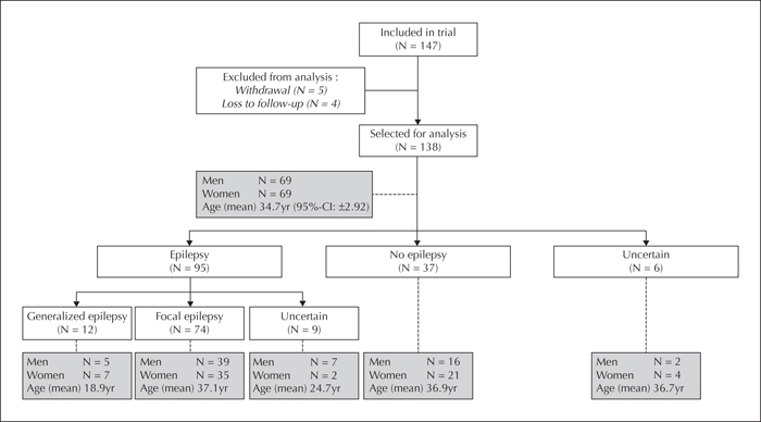
Aim. The diagnostic process for epilepsy can be lengthy and stressful, which may delay the start of treatment. The objective of this study was to determine the benefit of routine magnetoencephalography (MEG) with regard to diagnostic gain, compared to routine electroencephalography (EEG), EEG following sleep deprivation (EEGsd), and 24-hour EEG. Methods. In this prospective study, patients were included from two centres (Academic Centre for Epileptology Kempenhaeghe, Heeze and Elisabeth-Twee Steden Hospital, Tilburg) and MEG recording took place at a single centre (Amsterdam University Medical Centre, Vrije Universiteit Amsterdam) in The Netherlands. Consecutively referred patients from peripheral hospitals were included between August 2013 and March 2016. Patients were offered routine MEG in addition to EEG examination and MRI for the diagnosis of epilepsy. The final clinical diagnosis was based on all available clinical data and test results at the end of the diagnostic process. Sensitivity, specificity, and positive and negative predictive values were calculated for routine EEG, routine EEG plus additional EEG and MEG. In addition, diagnostic gain associated with MEG, relative to the other modalities, was calculated. Secondary outcome was congruence of localization of epileptiform discharges between MEG and MRI or final clinical diagnosis. Results. Based on a cohort of 138 patients, sensitivity and specificity was shown to be 31.6% and 78.4% for routine MEG, 31.6% and 100% for routine EEG, and 52.6% and 97.3% for routine EEG plus additional EEG, respectively. Routine MEG demonstrated a diagnostic gain of 16.8% compared to routine EEG and 9.5% compared to routine EEG plus additional EEG. In 35.7% of patients with a lesion on MRI that was consistent with the final clinical diagnosis, MEG showed epileptiform discharges in the same area.
Conclusion. Routine MEG may provide additional value during the initial diagnosis of epilepsy.
 Cette œuvre est mise à disposition selon les termes de la
Licence Creative Commons Attribution - Pas d'Utilisation Commerciale - Pas de Modification 4.0 International
Cette œuvre est mise à disposition selon les termes de la
Licence Creative Commons Attribution - Pas d'Utilisation Commerciale - Pas de Modification 4.0 International