Epileptic Disorders
MENUPresentation, diagnosis and treatment of bilateral Rasmussen's encephalitis in a 12-year-old female Volume 15, numéro 3, September 2013

epd.2013.0594
Auteur(s) : Katrina Peariso1 katrina.peariso@cchmc.org, Shannon M Standridge1, Barbara E Hallinan1, James L Leach2, Lili Miles3, Francesco T Mangano4, Hansel M Greiner1
1 Department of Neurology
2 Department of Radiology
3 Department of Pathology
4 Department of Neurosurgery, Cincinnati Children's Hospital Medical Center, Cincinnati, Ohio, USA
Correspondence: Katrina Peariso Department of Neurology, Cincinnati Children's Hospital Medical Center, Cincinnati, OH 45229, USA
Rasmussen's encephalitis (RE) is a syndrome of chronic localised encephalitis involving a single cerebral hemisphere, treatment of refractory epilepsy, and neurological impairment (Rasmussen et al., 1958). The aetiology of RE remains unknown. Neuropathological studies demonstrating neuroglial and lymphocytic responses consistent with other autoimmune CNS diseases (Pardo et al., 2004) and case reports demonstrating efficacy of rituximab therapy (Thilo et al., 2009) point to an autoimmune origin. Three clinical stages (prodromal, active, and residual) have been described and the syndrome is diagnosed based on published clinical criteria (Bien et al., 2005). However, atypical cases have been reported, including adolescent/adult onset, delayed epilepsy onset, absence of epilepsy, and extremely rare cases of bilateral RE (Andermann and Farrell, 2006; Bien et al., 2007; Ramesha et al., 2009; Guan et al., 2011). There have been a dozen reported cases of bilateral RE, but given the lack of biopsy/autopsy confirmation of bilateral involvement in the majority of cases, it is difficult to be certain of the diagnoses in all cases. Herein, we describe a case of biopsy-confirmed bilateral RE.
Case study
The patient was a 12-year-old, left-handed female who presented with new-onset seizures consisting of a painful left calf sensation that progressed to the contralateral leg and left arm with face numbness, followed by a “figure of four” sign related to tonic left arm extension and a bent right arm. The seizures lasted less than two minutes, and afterwards, she would have a left mouth droop and slurred speech for 20-30 minutes. Her neurological examination at presentation demonstrated right upper extremity dysmetria and intention tremor. Treatment with levetiracetam was initiated, but she continued to have intermittent breakthrough seizures. Her initial 24-hour EEG study was normal. Head CT and brain MRI obtained near the onset of her epilepsy showed marked volume loss and abnormal T2 signal in the left cerebral parenchyma, in addition to volume loss in the right cerebellum (figure 1B, C). Head CT had been obtained nearly five years prior to symptom onset after a minor head trauma (figure 1A), which showed milder right cerebellar volume loss, but no evidence of left hemispheric abnormality. Thus, the imaging findings at the onset of her epilepsy were consistent with interval left hemispheric volume loss and presumed cerebellar diaschisis.
The frequency and semiology of her seizures evolved over time. She was admitted six months after seizure onset for multiple daily seizures with interictal disinhibition, mood lability, and memory problems. The EEG performed during this hospitalisation captured multiple seizures, demonstrating right hemispheric onset in Pz-P4 (figure 2). Given her clinical presentation, a diagnosis of RE was considered in addition to infectious causes of encephalitis, progressive myoclonic epilepsy, mitochondrial epileptic encephalopathy, and an autoimmune-mediated epilepsy (table 1).
Table 1 Summary of the laboratory diagnostic work-up.
| Test | Results (where given) | Interpretation |
|---|---|---|
| Serum Amino Acids | Elevated Alanine | Likely secondary to seizure activity |
| Lactate Pyruvate |
1.60 mmol/L 0.12 mmol/L |
Normal |
| Carnitine | Total: 43.4 nmol/mL Free: 33.1 nmol/mL Short: 10.3 nmol/mL |
Normal |
| Acylcarnitine profile | Normal free carnitine Several longer chains Elevated 2 fold |
Fasting pattern |
| Ammonia | 21 μmol/L | Normal |
| Urine organic acids | Lactate elevated 2 fold | No diagnostic pattern |
| Urine metabolic screen | Normal | |
| TSH/Free T4/anti-TPO Ab | TSH: 1.07 mIU/mL FT4: 1.4 ng/dL Anti-TPO Ab: <5 IU/mL |
Normal |
| ESR | 5 mm/hr | Normal |
| CRP | <0.3 mg/dL | Normal |
| ANA | Negative | |
| Serum Lyme IgM/IgG | Negative | |
| Serum mycoplasma IgM/IgG | Negative | |
| Serum arboviral panel | Negative | |
| POLG1 | No known deleterious mutations | Negative |
| Progressive Myoclonic Epilepsy Panel (EPM1/2A/2B, Lafora, MERRF, EFHC1) |
No variants or mutations found | Negative |
| Muscle biopsy/ETC studies | No inclusions, RRF No ETC abnormalities |
Normal |
| CSF cell counts (WBC/RBC) | 0/71 | Normal |
| CSF glucose | 59 mg/dL | Normal |
| CSF protein | 39 mg/dL | Normal |
| CSF cultures and gram stain | No organisms; No growth | Normal |
| CSF OCB | 7 | Positive |
| CSF IgG | 2.6 mg/dL | Normal |
| CSF Amino Acids | Thr, Glu Elevated | Non-diagnostic pattern |
| CSF lactate/pyruvate | Normal | |
| CSF anti-NMDA | Negative | |
| CSF paraneoplastic antibody panel* | Negative | |
| CSF Arbovirus | Negative | |
| CSF VZV | Negative | |
| CSF HHV6 | Negative | |
| CSF HSV 1/2 | Negative | |
| CSF EBV | Negative | |
| CSF CMV | Negative | |
| CSF Enterovirus | Negative | |
| CSF Influenza A/B | Negative |
*Paraneoplastic antibody panel + Anti-NMDA Ab Mayo Clinic included antibodies against: NMDA, ANNA-1, ANNA-2, ANNA-3, AGNA-1, PCA-1, PCA-2, PCA-Tr, CRMP-5-IgG, PQ-type calcium channel, N-type calcium channel, ACh receptor, AChR ganglionic neuronal, neuronal (V-G) potassium channel, and striational Abs.
The patient's seizure frequency continued to increase, with a slightly more anterior localisation on EEG (figure 2). She developed a left hemiparesis, most severe in her distal left leg. Review of serial brain MRI over an eight-month period after seizure onset demonstrated increasing T2 hyperintensity in the left frontal and anterior parietal lobe and new increased T2 signal in the medial right frontal lobe (figure 1C, D, F). The left cerebral hemisphere and right cerebellar hemisphere volume loss remained stable. 18FDG PET showed intense glucose uptake in the parasagittal region of the right posterior frontal lobe near the central sulcus in the area of the new MRI signal, with otherwise extensive areas of decreased metabolism in most of the left frontal lobe, right cerebellar hemisphere, and the superior aspect of both parietal lobes (figure 1E). Interictal and ictal MEG source localisation supported a right parasagittal focus. With no obvious underlying aetiology and evolving clinical history, RE was the primary diagnostic consideration, however, the bilateral imaging findings complicated the diagnostic picture.
The patient's left leg and arm focal motor seizures evolved into EPC, in spite of therapeutic doses of multiple antiepileptic drugs (phenytoin, levetiracetam, oxcarbazepine, clonazepam, lacosamide, valproic acid, zonisamide, diazepam, vigabatrin, and phenobarbital), IVIg, and a four-week course of rituximab. A bicoronal craniotomy was performed and biopsies were taken from the parasagittal region of bilateral frontal pre-motor areas to aid in the diagnosis. Biopsy samples from both frontal cortices and white matter were consistent with a T-cell mediated encephalitis (figure 3). It was noted that the patient had been treated with rituximab, which effectively depletes CD20+ B cells, possibly altering some aspects of the biopsy sample. A neuropathological expert second opinion confirmed the diagnosis as most consistent with RE. With the patient's EPC and biopsy-confirmed chronic inflammatory changes, the criteria proposed by Bien and coworkers (Bien et al., 2005) for right hemispheric RE were met. Since the pathological process was clearly present in both hemispheres, she was diagnosed with bilateral RE.
Post-biopsy, trials of intravenous solumedrol, rituximab, and plasmapheresis did not provide meaningful improvement in the patient's symptoms. She was initiated on monthly intravenous immunoglobulin (IVIG) infusions. Functional MRI evaluation demonstrated left language lateralisation. The option of a right functional hemispherotomy to treat her EPC was discussed with the patient and her family. However, they were reticent to incur the expected neurological deficits associated with this procedure in the context of an uncertain prognosis for her left hemisphere, given that the biopsy demonstrated bilateral inflammation. They declined surgery at this time. Her seizure frequency has remained stable to mildly improved six months post biopsy, with parents reporting 6-15 seizures per day. She continues to have moderate left hemiparesis but is ambulatory and has use of her left hand, with stable cognitive function.
Discussion
This report describes the diagnostic challenges and treatment dilemmas that arise in a pathology-confirmed case of bilateral RE. Of the more than 200 reported cases of RE in over 50 years, there are less than 12 cases that meet the criteria for diagnosis of bilateral RE (Bien et al., 2005) (table 2), and only two have pathology-verified bilateral disease, performed at autopsy. This patient meets the two B-criteria described by Bien et al. (2005) for diagnosis of RE, namely epilepsia partialis continua and biopsy-confirmed T cell-dominated encephalitis with activated microglia and reactive astrogliosis. The patient presented with a right hemispheric syndrome of explosive right parasagittal seizures, interictal cortical dysfunction resulting in left hemiparesis, and EEG/MEG findings consistent with right parasagittal localisation. The patient did not demonstrate significant right hemispheric cerebral volume loss during the course of her illness, but it was unclear how much the aggressive immunomodulatory therapy she received would alter this finding. The pathology confirmed that the inflammation was clearly bilateral.
Table 2 Reported cases of bilateral Rasmussen's encephalitis.
| Reference | Age at first seizure |
Focal deficits |
EPC | Hemispheric atrophy MRI/CT |
Pathology confirmation |
|---|---|---|---|---|---|
| Guan et al., 2011 | 2 years | bilateral | + bilateral | + | + Left fronto-parietal; No pathology from right hemisphere |
| Chinchilla et al., 1994 | 5.9 years | bilateral | + bilateral | + | None |
| Chinchilla et al., 1994 | 3.5 years | unilateral | + bilateral | + | + single occipital lobe |
| Chinchilla et al., 1994 | 8.75 years | unilateral | + bilateral | + | + bilateral (necropsy) |
| Tobias et al., 2003 | 2.5 years | bilateral | No | + (bilateral) | + bilateral (necropsy) |
| Farrell et al., 2002 | 2.75 years | NR | + right | + | + right hippocampus |
| Silver et al., 1998 | 0.5 year | bilateral (L>R) | + bilateral | - | + right frontal |
| Silver et al., 1998 | 0.3 year | bilateral | + bilateral | + (bilateral) | None |
| Takahashi et al., 1997 | 0.17 years | bilateral | + bilateral | + (bilateral) | None |
| Takahashi et al., 1997 | 0.17 years | bilateral | + bilateral | + (bilateral) | None |
| Takahashi et al., 1997 | 2.4 years | bilateral | + bilateral | + (bilateral - left before right) | None |
| DeToledo and Smith, 1994 | 14 years | unilateral | + unilateral | NR | + left frontal lobe |
| DeToledo and Smith, 1994 | 11 years | unilateral | + unilateral | + | + bilateral |
This case report has some limitations. Although an extensive workup to investigate causes of encephalitis was performed, an undiagnosed aetiology cannot be ruled out. Second, to preserve function, restricted biopsies were obtained from both hemispheres, limiting the ability to diagnose a potential dual pathology, such as focal cortical dysplasia, that may have been possible with a larger resection. Third, although both hemispheres demonstrated similar pathology, the patient had no clear neurological symptoms from the progressive atrophy of the left cerebral hemisphere. This contrasts with previously reported cases of bilateral RE showing bilateral neurological signs (Chinchilla et al., 1994; Takahashi et al., 1997; Silver et al., 1998; Tobias et al., 2003; Guan et al., 2011). Nonetheless, there are reports of atypical cases of RE that have demonstrated diffuse hemiatrophy and functional impairment for 1.3-1.9 years prior to seizure onset in addition to cases without seizures (Bien et al., 2007).
The degree of hemiatrophy in the left cerebral hemisphere with crossed cerebellar diaschisis and minimal associated neurological dysfunction on initial examination is an unusual feature of this case. It seems unlikely that the patient would have had a chronic encephalitis involving the left cerebral hemisphere leading to crossed cerebellar diaschisis while remaining asymptomatic from a neurological and developmental perspective. In addition, the right cerebellar atrophy was noted prior to symptom onset, before the left hemisphere atrophy observed at epilepsy onset (figure 1A, B). Cianfoni et al. (2010) reported the presence of crossed cerebellar diaschisis on brain MRI at five years of follow-up in a patient with biopsy-confirmed RE. However, the timing of symptom onset with the corresponding imaging findings in this report does not correlate with that seen in our patient. The patient in our report was symptomatic for only one month before the left cerebral atrophy was first identified on MRI. Her parents reported a left-handed preference since 3 years of age, suggesting the left cerebral hemisphere volume loss and the crossed cerebellar diaschisis may be secondary to a remote injury, causing a pathological left-handedness. Cerebellar atrophy prior to hemispheric atrophy has been described after neonatal cerebellar injuries (Limperpoulos et al., 2005) in addition to cerebellar strokes in adults, although obvious areas of cerebellar injury were absent in imaging studies of this patient (figure 1A-C). Apparent evolution of left cerebral atrophy over four years argues against a completely static left hemispheric process. Various causes of primary pathology with secondary activation of inflammation, so-called “dual pathology” in RE, have been well-described (Silver et al., 1998). In this case report, dual pathology of our patient's left cerebral and right cerebellar atrophy can only be speculated.
The recommended treatment for seizure control in RE, based on expert opinion, is hemispherotomy. Immune modulatory therapy is a secondary treatment, mostly to help prevent the neurological decline observed in these patients. This treatment protocol is generally followed in patients with very early onset of disease and those in which the seizures are the most prominent morbidity in the illness. There is no such consensus for patients with bilateral disease. The treatment of our patient falls into a category described by Bien and Schramm (2009) as a therapeutic dilemma. This patient had a later clinical onset of disease suggesting less risk of mortality. Her acute syndrome was right hemisphere in origin, but she had clear bilateral cortical and white matter inflammatory changes on pathology. Post-hemispherotomy, she would be reliant on a compromised hemisphere leading to an uncertain prognosis. Bilateral RE treatment decisions should be approached on an individual basis, but aggressive immune therapy is a valid alternative to surgery.
Acknowledgements and disclosures
This work was not supported by any grant funding. This case report was presented as an abstract/poster at the 66th Annual Meeting of the American Epilepsy Society.
Shannon Standridge, DO is a speaker for Lundebeck pharmaceuticals. The remainder of the authors have no commercial or financial disclosures that could be construed as a conflict of interest.
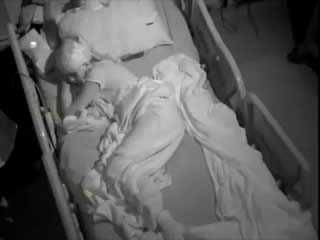
Legends for videosequences Representative seizure shown in the electroencephalograms of figure 2A, B, prior to development of left upper and lower limb epilepsia partialis continua. Key words for video research on www.epilepticdisorders.com Syndrome: Rasmussen syndrome Etiology: encephalitis Phenomenology: clonic seizure; tonic posture Localization: central (right)