Epileptic Disorders
MENUObstructive sleep apnea syndrome and nocturnal epilepsy with tonic seizures Volume 11, numéro 4, December 2009

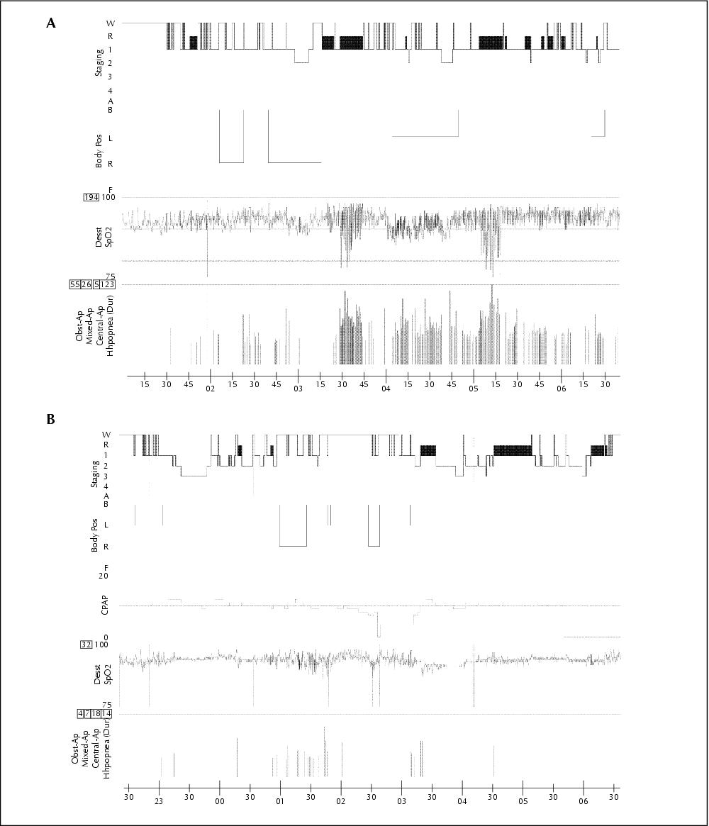
Figure 1 A) Diagnostic polysomnography. B) Polysomnography with CPAP treatment.W: wakefulness; R: REM; 1, 2, 3 and 4: sleep stages; Body pos: body position; B: supine; L: left lateral; R: right lateral; F: front; Desat: desaturations; the horizontal lines represent the levels of saturation: 100, 90 and 80%, respectively; Obst-Ap: obstructive apnea; Mixed-Ap: mixed apnea; Central-Ap: central apnea; Hypopnea (Dur): hypopneas; the height of the vertical lines represents the relative duration of respiratory events; the numbers within the squares represent the amount of desaturations and respiratory events; CPAP: the horizontal line represents the pressure of 10 mbar; horizontal axis: time in hours.