Epileptic Disorders
MENUIn the pursuit of the epileptogenic zone – listen carefully and look deeply Volume 23, numéro 4, August 2021

- Mots-clés : epilepsy, seizure semiology, stereo-electroencephalography, SEEG, electrocorticography, ECoG, epilepsy surgery
- DOI : 10.1684/epd.2021.1297
- Page(s) : 667-73
- Année de parution : 2021
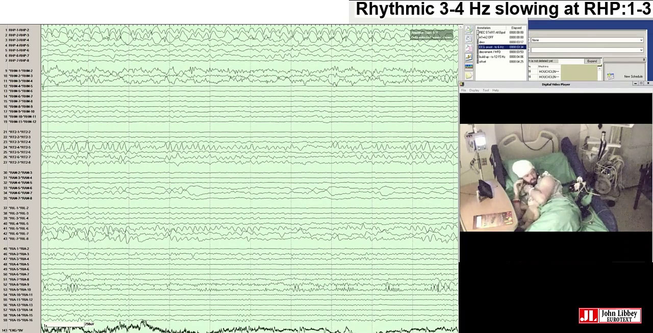
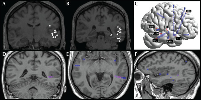
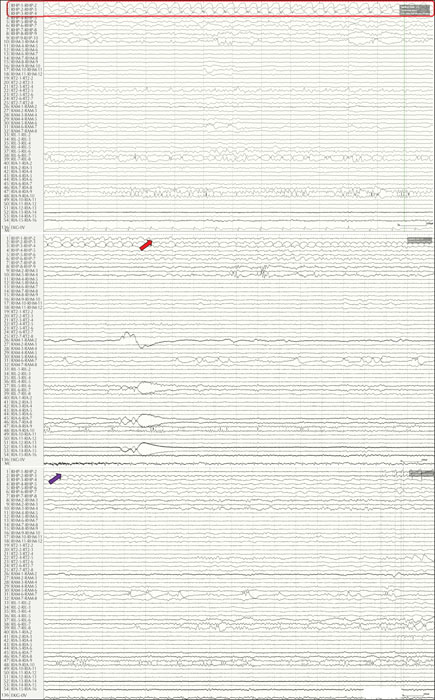
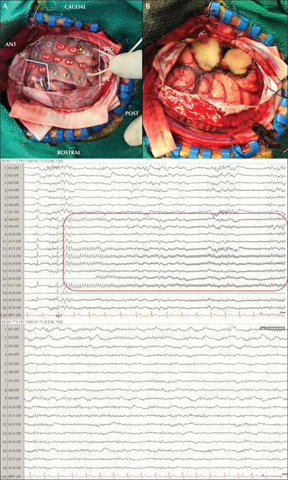
We report a 30-year-old right-handed man with a history of drug-resistant, non-lesional, childhood-onset focal epilepsy featuring (i) focal unaware seizures with left upper extremity automatisms and tonic posturing preceded by an aura of a ringing/beeping noise, and (ii) nocturnal hyperkinetic seizures. Non-invasive video-EEG, MEG, and PET were unable to delineate the epileptogenic zone (EZ) warranting an invasive investigation with bilateral depth electrodes (SEEG). SEEG data localized the EZ to the right superior temporal sulcus (STS) and right superior temporal gyrus (STG), wherein the auditory cortex lies, with subsequent ictal spread to anterior topography including the operculo-insular region. This hypothesis explained the patient's semiology consisting of focal aware seizures featuring auditory phenomena and nocturnal hyperkinetic seizures. Our multi-disciplinary team elected to proceed with a resection of the posterior right STG guided by electrocorticography (ECoG). Prior to resection, ECoG identified seizures arising from the peri-sylvian region, seemingly discordant to previous SEEG data. Following resection of the posterior right STG, ECoG continued to show seizures from contacts overlying the parietal operculum. It was not until the cortex was resected, at the depth of the right STS, that ECoG no longer showed epileptiform abnormalities. Pathology revealed focal cortical dysplasia type 1A.