Epileptic Disorders
MENUIctal atrial fibrillation during focal seizures: a case report and literature review Volume 21, numéro 3, June 2019

Figure 1
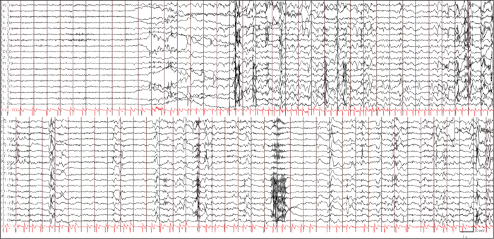
EEG tracing showing an electroclinical seizure arising from the left temporal lobe associated with a cardiac rhythm disorder. At seizure onset, a flattening of the background activity can be observed over the left anterior and middle temporal regions, followed by theta waves and then rhythmic sharp-wave activity, localized in the same regions; the ictal discharge eventually spreads to ipsilateral suprasylvian structures. ECG channel documents a seizure-related change in cardiac activity, consisting of paroxysmal atrial fibrillation, starting five seconds after seizure onset and ending 60 seconds after seizure termination. Technical details: 21-channel digital EEG recording with time-locked video and single-channel electrocardiography; electrodes were placed according to the 10 to 20 international system; bandpass filters 16 to 30 Hz; sensitivity 10 μV/mm.

Figure 2
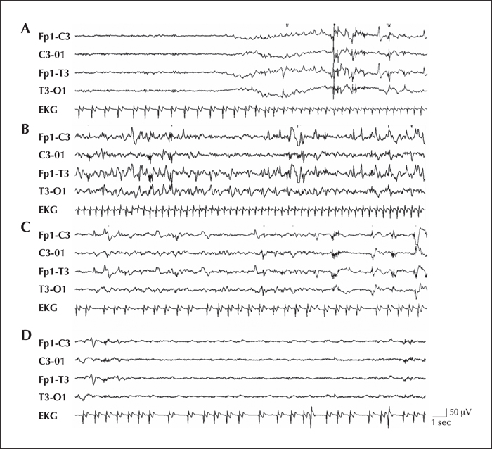
EEG samples emphasizing the ictal pattern and the related ECG changes during seizure evolution on the left temporal regions (A) EEG onset, (B) seconds 25-45, (C) seconds 50-70, (D) post-ictal phase.

