Epileptic Disorders
MENUElectropositive seizures and non-convulsive status epilepticus in a critically ill patient with prior skull defect Volume 22, numéro 2, April 2020

Figure 1
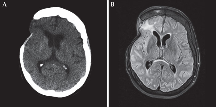
(A) Head CT without contrast demonstrating post-operative changes with skull defect and encephalomalacia in the right temporal and frontal lobes. (B) MRI T2 fluid attenuation inverse recovery (FLAIR) demonstrating known encephalomalacia and mild increased signal of the right insular cortex and posterior frontal operculum, felt to represent post-ictal changes.

Figure 2
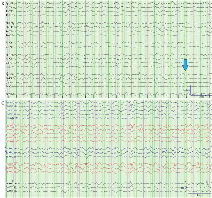
(A1) Typical interictal background with epoch revealing diffuse attenuation of the background activity with focal polymorphic theta activity over F4> F8 and Fz. Rare surface negative sharp waves are observed over F4>F8. (A2) Rare onset of a non-convulsive seizure characterized by fluctuating 1-Hz sharply contoured lateralized periodic discharges (LPDs) over F4 (most onsets did not feature LPDs), transitioning in the middle of the epoch to low-voltage irregular and arrhythmic fast activity with frequent intermixed low-amplitude positive spikes over F4 (arrow). (A3) Continuation of previous epoch of recording, highlighting an ongoing focal non-convulsive seizure, maximal over F4 and characterized by frequent intermixed surface positive low-amplitude spikes over F4 (arrow). (B) Typical ending of electrographic seizure activity with 1-2-Hz delta activity over F4, F8 (arrow). (C) Typical progression of a non-convulsive seizure viewed in a referential montage with surface positive sharp waves over F4 and F8. HFF: 70 Hz; LFF: 1 Hz ; 10 mm/sec, Notch off.

