Epileptic Disorders
MENUCortical surface intracranial electrodes identify clinically relevant seizures missed on scalp EEG after traumatic intracranial hemorrhage Volume 20, numéro 6, December 2018

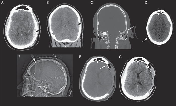
Figure 1
Pre- and post-operative CT imaging. Pre-operative CT head non-contrast (A) axial and (B) coronal slices showing left temporal epidural hematoma (arrows) with maximal radial diameter of 16 mm, (C) the overlying oblique non-displaced fracture of the left temporal bone (arrow), which extends through the squamous portion into the petrous portion (not shown), and (D) contralateral right convexity contracoup SDH (arrow). There was also a small amount of air tracking into the Eustachian tube and surrounding the fracture, but no disruption of the petrous carotid was visible on angiography (also not shown.) This resulted in mild effacement of the ambient cisterns and right greater than left lateral ventricles without significant midline shift. Note that this epidural hematoma crosses the suture lines and may be confused with an SDH, but this is due to the dura's skull insertion disruption by the fracture. The largest lesion was directly visualized above the dura intraoperatively, although smaller subdural fluid collections and intraparenchymal contusions were visible after opening dura. Immediately post-operative head CT non-contrast, after left hemicraniectomy and cortical electrode placement in the frontal lobe and estimated location of the motor cortex (arrows) (E) scout reconstruction, shows placement of cortical electrodes over the left frontal and parietal lobes, and (F) axial slice shows post-operative localization of the frontal lobe EEG lead (arrow: motor cortex lead not shown in this slice) and (G) the same slice windowed for soft tissue to show substantial improvement in mass effect after evacuation.

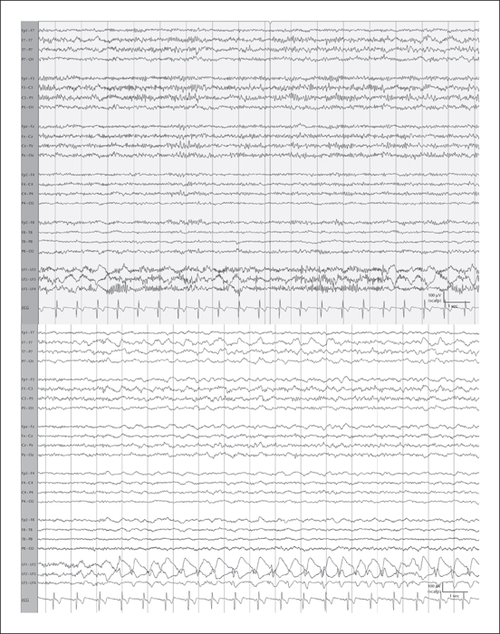
Figure 2
Scalp and frontal cortical IEEG and seizure. Upper panel: longitudinal bipolar montage scalp EEG showing diffuse slowing with overlying fast activity (12-20 Hz). Intermittent focal delta slowing and a breach rhythm are seen over the left hemisphere, maximal in the centrotemporal region. The bottom three bipolar channels display concurrent cortical IEEG derived from the frontal electrodes, as indicated by leads LF1-LF4. These demonstrate intermittent rhythmic delta activity that is at times sharply contoured. The non-recording parietal electrode channels are not shown. Lower panel: sharply contoured activity seen on cortical EEG has become rhythmic, following an initial evolution. There is spread to the LF3-LF4 channel, where a clear spike and wave morphology is seen. Waxing and waning rhythmic focal slowing without clear evolution is intermittently seen on concurrent scalp EEG despite persistence of the IEEG seizure. (Filter settings: LFF: 1 Hz; HFF: 70 Hz; notch: 60 Hz; sensitivity: 10 μV; timebase: 15 mm/sec.)

