Epileptic Disorders
MENUCharles Bonnet syndrome in hemianopia, following antero-mesial temporal lobectomy for drug-resistant epilepsy Volume 9, numéro 3, September 2007

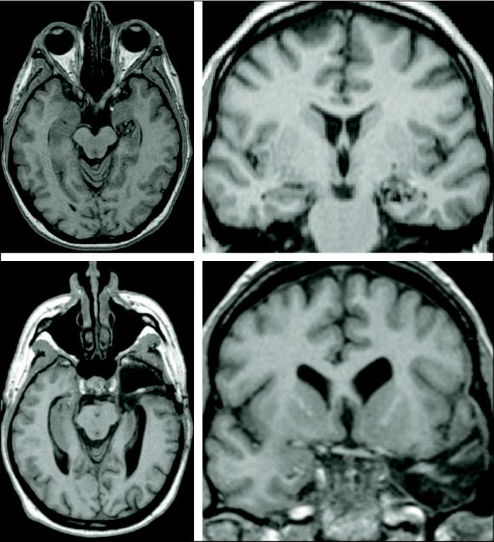
Figure 1 Upper panel: axial and coronal MRI images showing a cavernous angioma in the antero-mesial portion of the left temporal lobe. Lower panel: axial and coronal images showing the extent of the left antero-mesial temporal resection.
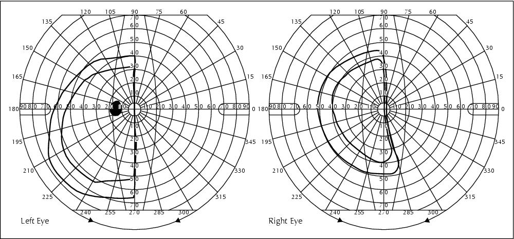
Figure 2 Post-operative visual field showing a right homonymous hemianopia.