Epileptic Disorders
MENUAn embarrassing aura Volume 21, numéro 3, June 2019

Figure 1
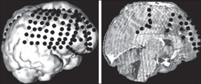
3-D representation of intracranial EEG electrode contacts: a 4 × 8 subdural grid was positioned over the dorsolateral frontal lobe as well as several (11) subdural strips to sample the ventromedial prefrontal cortex, the cingulate gyrus, the orbitofrontal, and frontopolar regions.

Figure 2
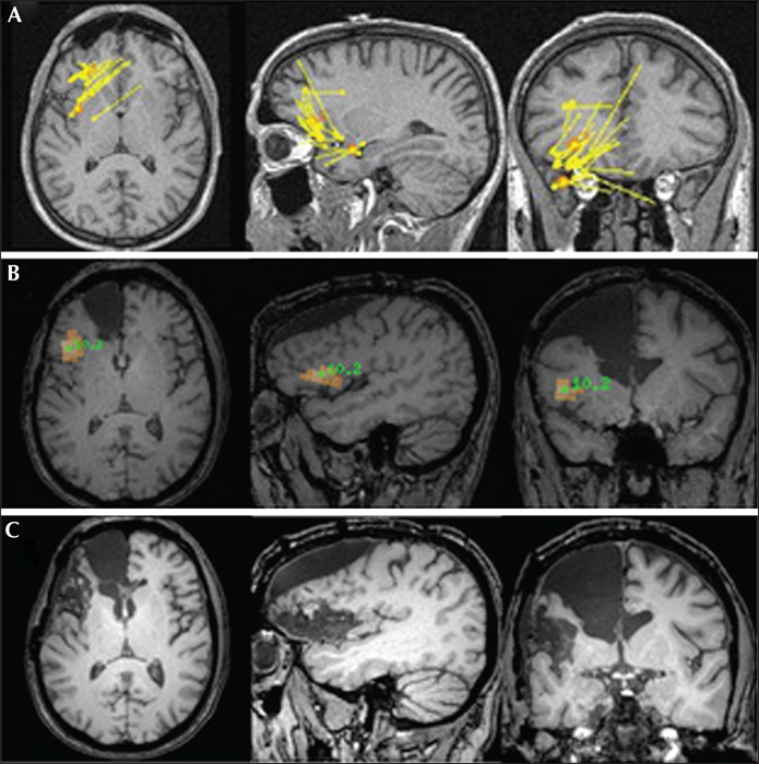
(A) Magnetoencephalography study (acquired prior to first surgery but analysed only after) showing clustered sources using the equivalent current dipole model over the anterior insula, posterior orbitofrontal cortex, and frontal operculum. (B) Magnetoencephalography study (performed after the first surgery) showing probable source of high-frequency gamma oscillations in the anterior insula using the Beamformer model. (C) MRI showing the location and extent of the first surgery over the anterior frontal region (arrow) and the second surgery at the junction of the frontal operculum, anterior insula, and posterior orbitofrontal cortex (triangle).

