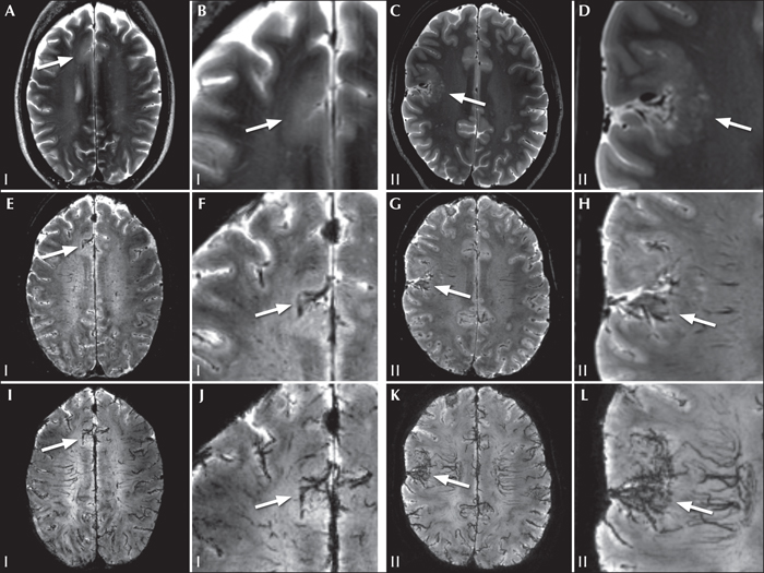
(A) MRI findings of patients with confirmed FCD and T2* signal changes; 7T T2 (A-D), T2* (E-H) and T2* minimum intensity projection (I-L) transverse reconstructions. Lesions are depicted in detail in (B), (D), (F), (H), (J) and (L). Patient I; FCD ILAE type IIb (A, B, E, F, I, J). No lesion was identified on 3T MRI and subtle grey-white matter junction blurring was seen on 7T T2weighted MRI (A, B). On T2* (E, F), the neighbouring sulcus appears to contain prominent vasculature. T2* minimum intensity projection (I, J) aids in the visual detection. Patient II; FCD ILAE type Ib (C, D, G, H, K, L). T2 -weighted MRI (C, D) shows grey-white matter junction blurring and cortical thickening indicative of FCD.In the same area, T2* (G, H) shows a wide sulcus with prominent vascular structures. T2* minimum intensity projection (K, L) strongly emphasizes the increased vasculature.
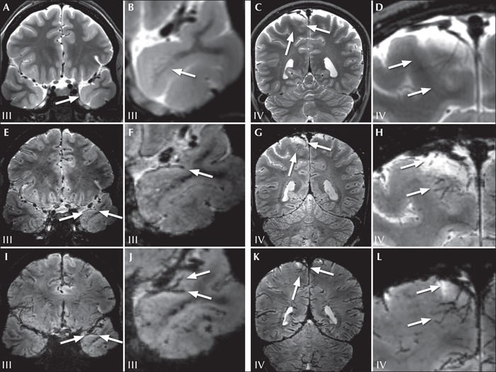
(B)MRI findings of patients with confirmed FCD and T2* signal changes. 7T T2 (A-D), T2* (E-H), and T2* minimum intensity projection (I-L) coronal reconstructions. Lesions are depicted in detail in (B), (D), (F), (H), (J) and (L). Patient III; mild malformation of cortical development type 2 (A, B, E, F, I, J). On T2 (A, B), blurring and subcortical hyperintensity represent developmental malformation. T2* (E, F) shows a wide Sylvian fissure but no clearly appreciable vascular changes. On T2* minimum intensity projection (I, J), there appears to be an increase in vascular signal in the superior temporal pole. Patient IV (C, D, G, H, K, L); FCD ILAE type IIb. On T2 (C, D), there is a notable large central parasagittal extracerebral space containing a large vein, but without evident dysplastic characteristics, however, on T2* (G, H), the large vein and smaller vasculature that drains from the dysplastic cortex (as confirmed by histological examination) is observed. (K, L) Image enhancement on T2* minimum intensity projection.
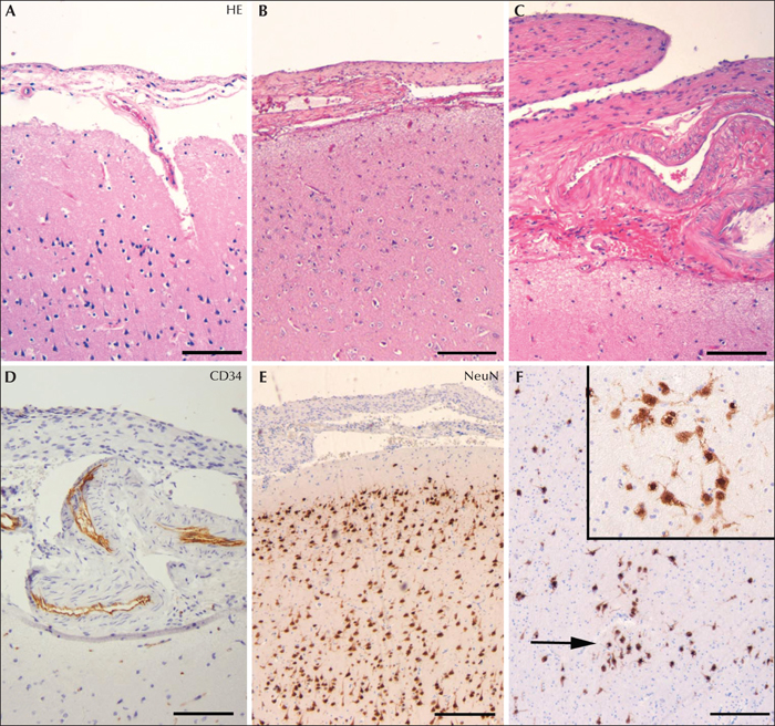
Histology. (A) Normal neocortex (HE: haematoxylin and eosin). (B-C) HE and (D) CD34 of neocortex from Patient III with thick (focally fibrotic) leptomeninges and prominent vascular structures. (E-F) NeuN of neocortex from Patient III without cortical dyslamination, but with microscopic neuronal clusters (arrow; insert in F) and excess of neurons of normal morphology in the deep white matter (isolated mild malformations of cortical development; mMCD type 2). Scale bar inA, B, E, F: 320 μm; C, D: 160 μm.
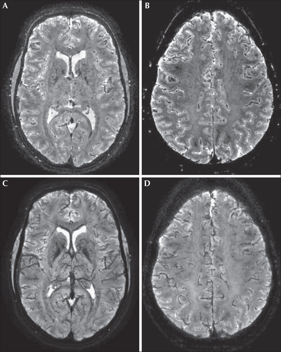
Transverse 7T T2* images of a healthy subject. T2*-weighted images showing transverse slices parallel to the anterior commissure-posterior commissure line at the level of the foramen of Monroe (A) and at the level of the cingulate sulcus (B). (A, B) Minimum intensity projections with 5-mm slab thickness at the level of (C) and (D), respectively. These images show the normal appearance of venous cerebral vasculature in a healthy subject.
Focal cortical dysplasia is one of the most common underlying pathologies in patients who undergo surgery for refractory epilepsy. Absence of a MRI-visible lesion necessitates additional diagnostic tests and is a predictor of poor surgical outcome. We describe a series of six patients with refractory epilepsy due to histopathologically-confirmed focal cortical dysplasia, for whom pre-surgical 7 tesla T2*-weighted MRI was acquired. In four of six patients, T2* sequences showed areas of marked superficial hypointensity, co-localizing with the epileptogenic lesion. 7 tesla T2* hypointensities overlying focal cortical dysplasia may represent leptomeningeal venous vascular abnormalities associated with the underlying dysplastic cortex. Adding T2* sequences to the MRI protocol may aid in the detection of focal cortical dysplasias.