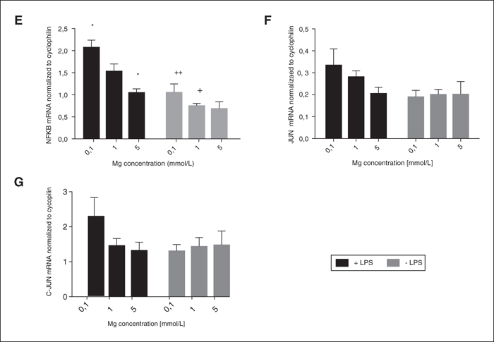
Effect of different concentrations of magnesium on NF-κB pathway gene expression. Data is shown as Mean ± SEM for n=4. * indicates P<0.05, compared to 1 mM MgSO4 in the same treatment (Dunnett's test), + indicates P<0.05, ++ indicates P<0.01 compared to LPS at the same magnesium concentration (t-test). A) Expression of TLR-4 mRNA. Two-way ANOVA showed significant interaction of magnesium concentration × presence of LPS (P=0.001). There was a significant effect of magnesium concentration (P=0.019) and LPS (P=0.039). B) Expression of IRAK1 mRNA. Two-way ANOVA showed no significant interaction of magnesium concentration × presence of LPS. C) Expression of TRAF6 mRNA. Two-way ANOVA showed no significant interaction of magnesium concentration × presence of LPS. D) Expression of IKB mRNA. Two-way ANOVA showed no significant interaction of magnesium concentration × presence of LPS. There was a significant effect of LPS (P=0.002). E) Expression of NF-ΚB 2 mRNA. Two-way ANOVA showed no significant interaction of magnesium concentration × presence of LPS. There was a significant effect of magnesium concentration (P<0.001) and LPS (P<0.001). F) Expression of JUN mRNA. Two-way ANOVA showed no significant interaction of magnesium concentration × presence of LPS. There was a significant effect of LPS (P= 0.04). G) Expression of C-JUN mRNA. Two-way ANOVA showed no significant interaction of magnesium concentration × presence of LPS. There was a significant effect of LPS (P= 0.043).
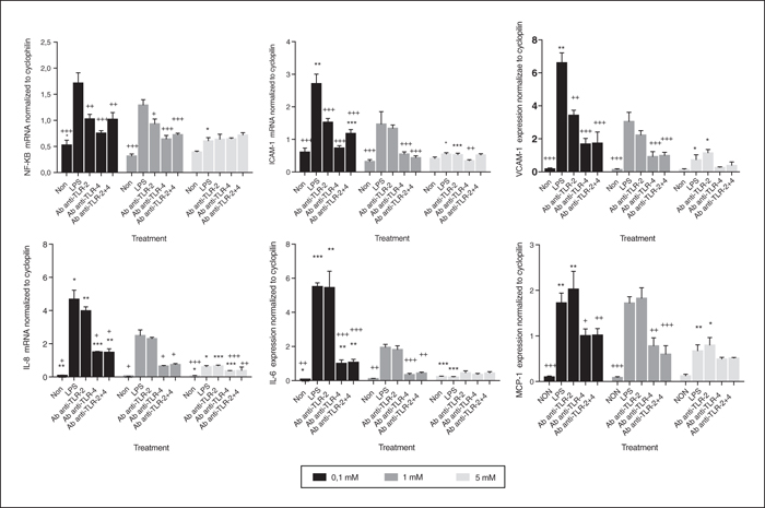
Effect of different concentrations of magnesium on HUVEC gene expression when treated or not treated with LPS and TLR antibodies. Data are shown as mean ± SEM for n=4. * indicates P<0.05, ** indicates P<0.01, *** indicates P<0.001 compared to 1 mM MgSO4 in same treatment (Dunnett t). + indicates P<0.05, ++ indicates P<0.01, +++ indicates P<0.001 compared to LPS at same magnesium concentration (Dunnett t). A) Expression of NF-κB mRNA. Two-way ANOVA showed a significant interaction of magnesium × blocker (P<0.001). There was a significant effect of magnesium concentration (P<0.001), and blocker (P<0.001). B) Expression of ICAM-1 mRNA. Two-way ANOVA showed a significant interaction of magnesium × blocker (P<0.001). There was a significant effect of magnesium concentration (P<0.001), and blocker (P<0.001). C) Expression of VCAM-1 mRNA. Two-way ANOVA showed a significant interaction of magnesium × blocker (P<0.001). There was a significant effect of magnesium concentration (P<0.001), and blocker (P<0.001). D) Expression of IL-8 mRNA. Two-way ANOVA showed a significant interaction of magnesium × blocker (P<0.001). There was a significant effect of magnesium concentration (P<0.001), and blocker (P<0.001). E) Expression of IL-6 mRNA. Two-way ANOVA showed a significant interaction of magnesium × blocker (P<0.001). There was a significant effect of magnesium concentration (P<0.001), and blocker (P<0.001). F) Expression of MCP-1 mRNA. Two-way ANOVA showed a significant interaction of magnesium × blocker (P=0.005). There was a significant effect of magnesium concentration (P<0.001), and blocker (P<0.001).
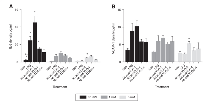
Effect of different concentrations of magnesium on HUVECs protein concentration treated or not with LPS and TLR antibodies. Data are shown as mean ± SEM for n=4. * indicates P<0.05 compared to 1mM MgSO4 in same treatment (Dunnett t). + indicates P<0.05, ++ indicates P<0.01 compared to LPS at same magnesium concentration (Dunnett t). A) Protein concentration of IL-8. Two-way ANOVA showed a significant interaction of magnesium × blocker (P<0.001). There was a significant effect of magnesium concentration (P<0.001), and blocker (P<0.001). B) Protein concentration of VCAM-1. Two-way ANOVA showed no significant interaction of magnesium × blocker. There was a significant effect of magnesium concentration (P<0.001), and blocker (P<0.001).
The aims of this study were to determine whether low concentrations of magnesium in vitro exacerbated the human umbilical vein endothelial cell (HUVEC) response to inflammatory challenge, and whether expression of the nuclear factor kappa-light-chain-enhancer of activated B cells (NF-κB) through the toll-like receptor 4 (TLR4) played a role in this process. HUVECs were incubated with different concentrations of Mg (low- 0.1mM, control- 1mM, high- 5mM) for 72 h before being stimulated with bacterial lipopolysaccharide (LPS) for 4 h. The response of cells to LPS was greater in cells cultured in low Mg, relative to control cells and suppressed in high Mg. Expression of NF-κB was increased in low-Mg and decreased with high Mg. Low Mg increased the expression of TLR4 mRNA, but only in the presence of LPS. Antibody blockade of TLR4 but not TLR2 blunted the response of cells to LPS in low Mg, such that they were similar to unblocked 1mM Mg cells. Associations of Mg with cardiovascular disease may therefore relate to inflammatory responses mediated through the TLR4/NF-κB pathway.